
Practice question
Question
Variant detection by cytological studies involves:
Explanation
Cytological analysis visualizes nuclear integrity to uncover ploidy changes and structural rearrangements. Actively growing root tips or callus are pretreated with mitotic inhibitors such as colchicine or paradichlorobenzene to accumulate metaphases, fixed in Carnoy's fluid, hydrolyzed with HCl, and stained with acetocarmine, Feulgen reagent, or fluorescent DAPI binding AT-rich DNA. Squash preparations allow chromosome counting, measurement of arm ratios, and identification of deletions, duplications, translocations, and fragments under light or epifluorescence microscope. Monitoring lagging chromosomes, multipolar spindles, and nuclear fragmentation reveals instability induced by 2,4-D and aging cultures. Flow cytometry supplements microscopy by quantifying DNA content quickly. In contrast techniques like PCR or ELISA detect sequence or protein but not chromosome behavior. Hence staining and microscopy remains central for cytological variant detection in somaclonal screening programs aimed at maintaining euploid stability. Preparation includes pretreatment with 8-hydroxyquinoline, fixation in acetic ethanol, and enzyme maceration to spread chromosomes. Analysis of karyotype asymmetry and satellite association identifies subtle translocations. This direct visualization complements molecular markers, ensuring detection of large-scale genome rearrangements that PCR alone cannot reveal in somaclonal screening.
Related blogs
Guides and insights connected to this topic
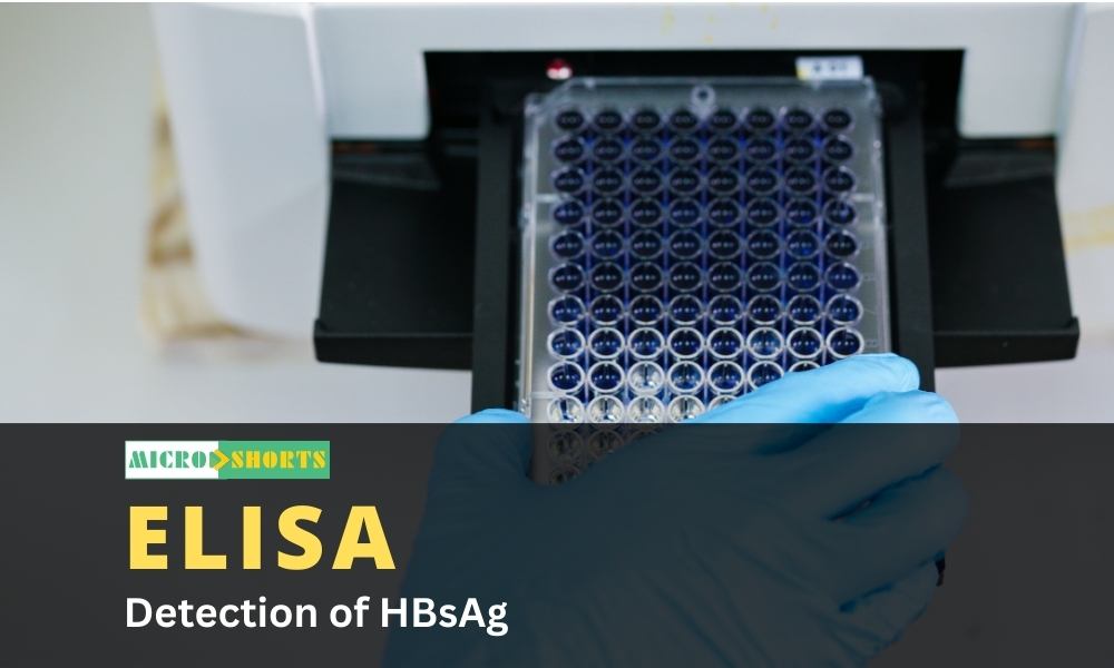
ELISA Detection of HBsAg
Introduction Hepatitis B or Serum Hepatitis is one of the most common infectious diseases in the world and has become a global public health...

Foodborne Giardiasis- Food Poisoning by Giardia species
Introduction Parasites are widely spread in the environment and are responsible for causing diseases associated with diarrheal illness. Food...

Wheatley Trichrome Staining
Wheatley Trichrome Staining definitionWheatley Trichrome staining technique is a special permanent satin used in parasitology for the detect...
Discussion
Comments
Please log in to join the discussion.
Login to commentNo comments yet. Be the first to start the discussion.