
Practice question
Question
Which microscopy is ideal for co-localization studies of two fluorophores?
Explanation
Related blogs
Guides and insights connected to this topic
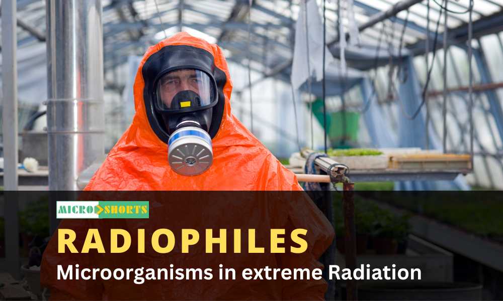
Microorganisms in extreme Radiation (Radiophiles)
Radiophile Definition and CharacteristicsRadiophiles are a group of extremophiles that are capable of surviving extreme forms of radiations...
Experiment 16: Flash Cards / Models / Specimens Showing Examples of Homologous and Analogous Organs
AIM To study homology and analogy with the help of models/charts of animals and models/charts/specimens of plants. PRINCIPLE Homologous organs are functionally dissimilar but anatomically or structurally similar. Different modes of life have modified the organs to enable survival...
Experiment 15: Identification and Study of Symbiotic Associations (Root Nodules of Leguminous Plants, Lichens) and Parasitism (Cuscuta on Host)
AIM To observe model specimen showing symbiotic association in root nodules of leguminous plants, Cuscuta on host, lichens. PRINCIPLE Symbiosis is close association between two different organisms where at least one is benefited. It includes mutualism where both are benefited, pa...

Discussion
Comments
Please log in to join the discussion.
Login to commentNo comments yet. Be the first to start the discussion.