
Structure of Lassa Virus
- The
virus is a single-stranded RNA virus (RNA virus – virus
whose genetic material is RNA, not DNA) belonging to the virus family Arenaviridae
(– a family of enveloped RNA viruses).
- The
virion is spherical particles (virion – complete infectious
virus particle) with an average diameter of 90–110 nm (nanometer
– very small unit of length).
- Lassa
virus is a single-stranded RNA virus that is enveloped in lipid (–
surrounded by a lipid membrane derived from host cell) with glycoprotein
spikes (– protein molecules with sugar groups attached)
protruding from the outside surface.
- Glycoproteins on the surface of the virion form T-shaped spikes (– spike-like projections) extending 7–10 nm from the envelope.
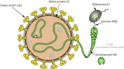
Figure: Structure of Lassa Virus, Source: Swiss Institute of Bioinformatics
Genome of Lassa Virus
- It
contains two species of RNA (– two separate RNA segments)
called the small and large units, and each unit has two genes at
opposite ends that do not overlap (– ambisense genome organization).
- The
small unit has some double stranded areas (– regions where RNA
pairs with itself) that form stem-loop structures (– folded
RNA structures important for regulation).
- The
large species of RNA encodes for the Z protein (– zinc-binding
regulatory protein) and L protein (– RNA-dependent RNA
polymerase) at the 5’ and 3’ ends (– ends of RNA strand)
respectively, and the small species of RNA encodes for glycoprotein
and nucleoprotein (– protein that binds viral RNA) at the 5’
and 3’ ends respectively.
- Lassa
virus consists of four lineages (– genetically distinct groups),
which have a strain variation of 27% in nucleotides and 15% in amino
acids (– genetic and protein-level variation).
- The
large segment encodes a small zinc-binding protein (Z) (–
protein involved in viral regulation) that regulates transcription
(– synthesis of RNA) and replication (– copying of viral
genome) and the RNA polymerase (L) (– enzyme that
synthesizes RNA).
- The
small segment encodes the nucleoprotein (NP) (– RNA-binding
protein) and the surface glycoprotein precursor (GP) (–
inactive form of spike protein), also known as the viral spike,
which is proteolytically cleaved (– cut by enzymes) into the
envelope glycoproteins GP1 and GP2 that bind to the alpha-dystroglycan
receptor (– host cell surface receptor) and mediate host
cell entry (– virus entering the cell).
- The
gene that encodes for the nucleoprotein is 1,710 nucleotides long,
and the protein has 569 amino acids (– building blocks of
proteins).
- The
gene that encodes for the glycoprotein is 1,473 nucleotides long.
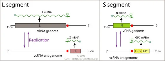
Figure: Genome of Lassa Virus, Source: Swiss Institute of
Bioinformatics
Epidemiology of Lassa Virus
- The
Lassa virus is so named because, in 1969, it was first isolated and
correlated as the causative agent (– disease-causing agent) of Lassa
fever in a small town called Lassa in North-eastern Nigeria.
- Lassa
fever is endemic (– constantly present in a region) in parts
of West Africa including Sierra Leone, Liberia, Guinea, and
Nigeria; however, other neighboring countries are also at risk, as the
animal vector (– organism that transmits disease) for Lassa
virus, the multimammate rat (Mastomys natalensis) (– natural
reservoir host), is distributed throughout the region.
- Lassa
virus consists of four lineages, three of these lineages are
located in Nigeria, while the other can be found in Guinea,
Liberia, and Sierra Leone.
- The
number of Lassa virus infections per year in West Africa is
estimated at 100,000 to 300,000, with approximately 5,000 deaths.
- In
some areas of Sierra Leone and Liberia, it is known that 10%–16%
of people admitted to hospitals every year have Lassa fever, which
indicates the serious impact (– high disease burden) of the
disease on the population of this region.
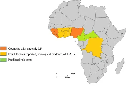
Figure: Epidemiology of Lassa Virus, Source: CDC
Transmission of Lassa Virus
- Humans
contract the virus primarily through contact with the contaminated
excreta (– urine or feces containing virus) of Mastomys
natalensis (– multimammate rat, natural host) rodents, which is
the natural reservoir (– organism that harbors the virus)
for the virus.
- The
virus is transmitted to humans through cuts and scratches (–
breaks in skin) or inhaled via dust particles (– airborne
transmission) in the air.
- Secondary
transmission (– human-to-human spread) of the virus between
humans occurs through direct contact with infected blood or bodily
secretions (– fluids released from the body).
Replication of Lassa Virus
- The
Lassa virus gains entry into the host cell by means of the cell-surface
receptor alpha-dystroglycan (alpha-DG) (– host receptor protein),
a versatile receptor for proteins of the extracellular matrix (–
structural network outside cells).
- After
virus enters the cell by alpha-dystroglycan mediated endocytosis (–
receptor-driven uptake into cell), a low-pH environment (–
acidic condition) triggers pH-dependent membrane fusion (–
fusion of viral and host membranes) and releases the RNP complex
(viral ribonucleoprotein – viral RNA bound to proteins) into
the cytoplasm (– fluid part of the cell).
- Viral
RNA is unpacked, and replication and transcription (– genome
copying and RNA synthesis) initiate in the cytoplasm.
- As
replication starts, both S and L RNA genomes (– small and large
viral RNA segments) synthesize the antigenomic S and L RNAs (–
complementary RNA copies), and from the antigenomic RNAs, genomic S
and L RNAs are synthesized.
- Both genomic
and antigenomic RNAs (– original and complementary strands) are
needed for transcription and translation (– RNA and protein
synthesis).
- S
RNA encodes GP (– glycoprotein) and NP (viral
nucleocapsid protein – RNA-binding protein), and L RNA
encodes Z (– regulatory protein) and L proteins (–
RNA polymerase).
- The primary
transcription (– first round of mRNA synthesis) first
transcribes mRNAs (– messenger RNAs) from the genomic S
and L RNAs, which code NP and L proteins, respectively.
- Transcription
terminates (– stops) at the stem-loop (SL) structure (–
folded RNA structure) within the intergenomic region (–
region between genes).
- Arenaviruses
(– virus family of Lassa virus) use a cap-snatching strategy
(– stealing caps from host mRNA) to gain cap structures (–
protective RNA modifications) from the cellular mRNAs, mediated by the
endonuclease activity of the L polymerase (– RNA-cutting
function) and the cap-binding activity of NP (– cap
recognition by nucleoprotein).
- Antigenomic
RNA transcribes viral genes GPC and Z, encoded in genomic
orientation (– gene direction), from S and L segments,
respectively.
- After
translation of GPC (– glycoprotein precursor), it is post-translationally
modified (– chemically altered after synthesis) in the endoplasmic
reticulum (– protein processing organelle).
- GPC
is cleaved (– cut enzymatically) into GP1 and GP2 at the
later stage of the secretory pathway (– protein transport system).
- Cleaved glycoproteins (– processed spike proteins) are incorporated into the virion envelope (– viral outer membrane) when the virus buds and releases (– exits the cell) from the cell membrane.
Pathogenesis of Lassa Virus
- When
initiating an infection, the Lassa virus attaches to a receptor on the
cell surface with the glycoprotein GP-1 (– attachment protein).
- Its
initial sites of replication include dendritic cells (DC) (–
antigen-presenting immune cells) and macrophage–monocyte cells
(– immune cells involved in phagocytosis), and it is then delivered
throughout the entire body.
- Infected
DC fail to secrete proinflammatory cytokines (– immune
signaling molecules), do not upregulate costimulatory molecules
(– signals needed for T-cell activation) such as CD40, CD80, and
CD86, and poorly induce proliferation of T cells (–
multiplication of immune cells).
- Lassa
virus prevents a host’s innate immune system (– first line of
immune defense) by NP activity (– immune-suppressive action
of nucleoprotein).
- Patients
infected with LASV (– Lassa virus) produce IgM and IgG
antibody isotypes (– early and long-term antibodies).
- Neutralizing
antibodies (– antibodies that block infection) appear months
after acute infection (– initial severe phase) is resolved,
and the titers (– antibody concentration) are often low.
- The
neutralizing antibody titers continue to rise even several months after convalescence
(– recovery period) has been established, which may indicate constant
stimulation of B cells (– antibody-producing cells) due to low
levels of virus persistence (– remaining virus in body).
- Antibodies
in seroconverted individuals (– people who developed antibodies)
are specific to GPC, NP, and, likely, Z protein (– viral
regulatory protein).
Clinical Manifestations of Lassa Virus
- The incubation
period (– time between infection and symptoms) of Lassa
fever ranges from 6–21 days.
- The spectrum
of clinical effects (– range of disease severity) manifested in
Lassa fever ranges from asymptomatic (– without symptoms)
infection to multi-organ system failure (– failure of multiple
organs) and death.
- The onset
of the disease, when it is symptomatic, is usually gradual,
starting with fever, general weakness, and malaise (–
general feeling of discomfort).
- After a few days, headache, sore throat, muscle pain, chest pain, nausea, vomiting, diarrhea, cough, and abdominal pain may follow.
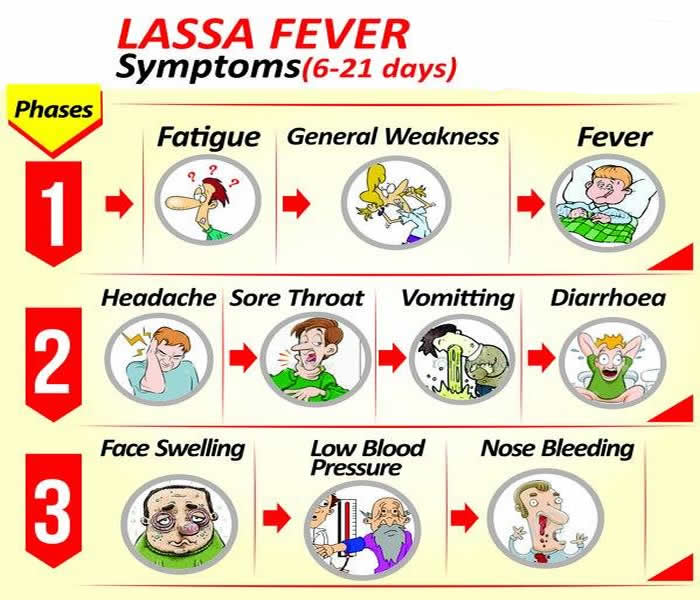
- In severe
cases, facial swelling, fluid in the lung cavity (–
pleural effusion), bleeding from the mouth, nose, vagina or
gastrointestinal tract (– hemorrhagic manifestations), and low
blood pressure (– hypotension) may develop.
- Shock
(– circulatory failure), seizures (– abnormal electrical
brain activity), tremor (– involuntary shaking), disorientation
(– confusion), and coma (– loss of consciousness) may
be seen in the later stages.
- Death
from Lassa fever most commonly occurs 10 to 14 days after symptom onset.
Diagnosis of Lassa Virus
- Detection
of IgM and IgG antibodies (– early and long-term immune
antibodies) as well as Lassa antigen (– viral protein)
by enzyme-linked immunosorbent serologic assays (ELISA) (–
antibody-based diagnostic test).
- The
virus can be uncovered using reverse transcription PCR (RT-PCR) (–
molecular method to detect viral RNA).
- Virus
isolation by cell culture, however, this procedure should only be done
in a high-containment laboratory (– biosafety level laboratory)
with good laboratory practices (– safety protocols). Mice
and guinea pigs have been evaluated as models of LASV infection
(– experimental animals for study).
- Immunohistochemistry
(– antibody-based tissue staining method), performed on formalin-fixed
tissue specimens (– preserved tissues), can be used to make a post-mortem
diagnosis (– diagnosis after death).
Treatment of Lassa Virus
- Ribavirin
(– antiviral drug) is only effective if administered early in
infection, within the first 6 days after disease onset.
Prevention and Control of Lassa Virus
- No
vaccine (– preventive immunization) for Lassa fever is
currently available for use in humans.
- Prevention
by promoting good community hygiene (– cleanliness practices at
community level) to discourage rodents (– disease-carrying
animals) from entering homes.
- Effective
measures include storing grain and other foodstuffs in rodent-proof
containers, disposing of garbage far from the home, maintaining
clean households, and keeping cats (– rodent control).
- Avoiding
contact with blood and body fluids (– infection source) while
caring for sick persons.
- In health-care settings, staff should always apply standard infection prevention and control precautions (– universal safety measures) when caring for patients, regardless of their presumed diagnosis. These include basic hand hygiene, respiratory hygiene, use of personal protective equipment (– PPE such as gloves and masks), safe injection practices, and safe burial practices.