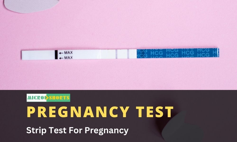
Principle
One step pregnancy
test utilize the principle of Immunochromatography, A unique two site
immunoassay on a membrane. As the test sample flows through the membrane
assembly of the dipstick, the colored anti-hcG-colloidal gold conjugate
complexes with the hcG in the sample.
This complex moves
further on the membrane to the test region where it is immobilized by the
anti-hcG coated on the membrane leading to formation of a purple colored band
which confirm a positive test result. Absence of this colored band in the test
region indicate a negative test result.
The unreacted
conjugate and unbound complex if any move further on the membrane and are
subsequently immobilized by the anti-rabbit antibodies coated on the membrane
at the control region, forming a purple band. This control band serves to
validate the test results.
Specimen: urine sample
Requirements
- Each individual
pouch contains.
- DIPSTICK:
membrane assembly Predispensed with anti-HCG antiserum and anti-rabbit
antiserum at the respective regions.
- Desiccant pouch.
- Package insert.
Procedure
- Collect urine
specimen in a clean test tube. Ensure that only sufficient quantity of the
specimen is collected to allow submerging the red area of the dipstick.
- Bring the sealed
pouch to room temperature, open the pouch and remove the dipstick. Once opened,
the dipstick must be used immediately.
- Dip the red area of
the dipstick in the urine specimen submerging only the red area.
- Observe for the
release of the colloidal gold complex on the membrane. This would be seen as a
purple moving front on the membrane and could take 10 to 15 seconds to appear
depending upon the sample.
- Remove the dipstick
and place horizontally on a flat surfaces Alternatively the dipstick may be
left to stand in the specimen for the entire duration of the test ensuring only
the red area is left submerged in the specimen.
- At the end of five
minutes read the results as follows
Clinical significance
Human chronic
gonadotropin (hcG), a glycoprotein hormone secreted by viable placental tissue
during pregnancy, is excreted in urine approximately 20 days after the last
menstrual period. The levels of hcG rise rapidly reaching peak levels after
60-80 days. The appearance of hcG in urine soon after conception and its rapid
rise in concentration makes it an ideal marker for the early detection and
confirmation of pregnancy.
However elevated hcG
levels are frequently associated with trophoblastic and non-trophoblastic
neoplasms and hence these condition should before a diagnosis of pregnancy can
be made. One step pregnancy test detects the presence of hcG in urine specimen,
qualitatively, at concentration as low as 10mlU/ml in less than five minutes.
Reference
KIT USED
– ORCHID BIOCHEMICAL SYSTEM
CLUE HCG


