Introduction
Having been
constructed in the 16th Century, Microscopes have revolutionalized science with
their ability to magnify small objects such as microbial cells, producing
images with definitive structures that are identifiable and characterizable.
What are Microscopes?
Microscopes
are instruments that are used in science laboratories to visualize very
minute objects such as cells, and microorganisms, giving a contrasting image that
is magnified. Microscopes are made up of lenses for magnification, each with
its own magnification powers. Depending on the type of lens, it will magnify
the specimen according to its focal strength.
Their ability
to function is because they have been constructed with special components that
enable them to achieve high magnification levels. They can view very small
specimens and distinguish their structural differences, for example, the view
of animal and plant cells, viewing microscopic bacterial cells.
Microscopes
are generally made up of structural parts for holding and supporting the
microscope and its components and the optical parts which are used for
magnification and viewing of the specimen images. This description defines the
parts of a microscope and the functions they perform to enable the
visualization of specimens.
Structural parts of a microscope and their functions
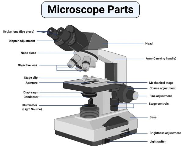
There are
three structural parts of the microscope i.e. head, base, and arm.
- Head – This is also known as the body.
It carries the optical parts in the upper part of the microscope.
- Base – It acts as microscopes support.
It also carries microscopic illuminators.
- Arms – This is the part connecting the
base and to the head and the eyepiece tube to the base of the microscope.
It gives support to the head of the microscope and it is also used when
carrying the microscope. Some high-quality microscopes have an articulated
arm with more than one joint allowing more movement of the microscopic
head for better viewing.
Optical parts of a microscope and their functions
The optical
parts of the microscope are
used to view, magnify, and produce an image from a specimen placed on a slide.
These parts include:
- Eyepiece – also known as the ocular.
This is the part used to look through the microscope. Its found at the top
of the microscope. Its standard magnification is 10x with an optional
eyepiece having magnifications from 5X to 30X.
- Eyepiece tube – it’s the eyepiece
holder. It carries the eyepiece just above the objective lens. In some
microscopes such as the binoculars, the eyepiece tube is flexible and can
be rotated for maximum visualization, for variance in distance. For
monocular microscopes, they are none flexible.
- Objective lenses – These are the major
lenses used for specimen visualization. They have a magnification power of
40x-100X. There are about 1- 4 objective lenses placed on one microscope,
in that some are rare facing and others face forward. Each lens has
its own magnification power.
- Nose piece – also known as the
revolving turret. It holds the objective lenses. It is movable hence it
cal revolve the objective lenses depending on the magnification power of
the lens.
- The Adjustment knobs – These are knobs
that are used to focus the microscope. There are two types of adjustment
knobs i.e fine adjustment knobs and coarse adjustment knobs.
- Stage – This is the section in which
the specimen is placed for viewing. They have stage clips that hold the
specimen slides in place. The most common stage is the mechanical stage,
which allows the control of the slides by moving the slides using the
mechanical knobs on the stage instead of moving them manually.
- Aperture – This is a hole on the microscope stage,
through which the transmitted light from the source reaches the stage.
- Microscopic illuminator – This is the microscopes
light source, located at the base. It is used instead of a mirror. It captures
light from an external source of a low voltage of about 100v.
- Condenser – These are lenses that are
used to collect and focus light from the illuminator into the specimen.
They are found under the stage next to the diaphragm of the microscope.
They play a major role in ensuring clear sharp images are produced with a
high magnification of 400X and above. The higher the magnification of the
condenser, the more the image clarity. More sophisticated microscopes come
with an Abbe condenser that has a high magnification of about 1000X.
- Diaphragm – it’s also known as the
iris. Its found under the stage of the microscope and its primary role is
to control the amount of light that reaches the specimen. It’s an
adjustable apparatus, hence controlling the light intensity and the size
of the beam of light that gets to the specimen. For high-quality
microscopes, the diaphragm comes attached with an Abbe condenser and
combined they are able to control the light focus and light intensity that
reaches the specimen.
- Condenser focus knob – this is a knob that moves
the condenser up or down thus controlling the focus of light on the specimen.
- Abbe Condenser – this is a condenser specially
designed for high-quality microscopes, which makes the condenser to be
movable and allows very high magnification of above 400X. High-quality
microscopes normally have a high numerical aperture than objective lenses.
- The rack stop – It controls how far the stages
should go preventing the objective lens from getting too close to the
specimen slide which may damage the specimen. It is responsible for
preventing the specimen slide from coming too far up and hitting the
objective lens.
Parts of a Microscope Revision Questions (FAQs)
Q. Define
a Microscope.
Ans.
Microscopes are
instruments that are used in science laboratories, to visualize very minute
objects such as cells, and microorganisms, giving a contrasting image, that is
magnified.
Q. State
functions of a microscope.
Ans. A microscope is usually used for the study of microscopic algae, fungi, and biological specimens.
Q.
Differentiate between a condenser and an Abbe condenser.
Ans. Condensers are
lenses that are used to collect and focus light from the illuminator into the
specimen. They are found under the stage next to the diaphragm of the
microscope. They play a major role in ensuring clear sharp images are produced
with a high magnification of 400X and above. Abbe condenser is
a condenser specially designed for high-quality microscopes, which makes the
condenser to be movable and allows very high magnification of above 400X.
High-quality microscopes normally have a high numerical aperture than objective
lenses.
Q. What is
the magnification power of the objective lenses?
Ans. Objective lenses have
a magnification power of 40X to 100X.
Q. How
does the eyepiece compare to the objective lens?
Ans. The eyepiece, also known
as the ocular is the part used to look through the microscope. Its found at the
top of the microscope. Its standard magnification is 10x with an optional
eyepiece having magnifications from 5X – 30X. Objective Lens are
the major lenses used for specimen visualization. They have a magnification
power of 40x-100x. There are about 1- 4 objective lenses placed on one
microscope, in that some are rare facing and others face forward.
Q. Why is
the rack stop included in the microscope from the factory, and can it be
replaced?
Ans. Rack stop is
included in the microscope for preventing the specimen slide from coming too
far up and hitting the objective lens.
Q. What is
a magnification power?
Ans. Magnification of a lens is
defined as the ratio of the height of an image to the height of an object.
Microscope magnification measures the total enlargement of the image of an
object. Magnification power is the product of eyepiece lens power and objective
lens power.
Q. Differentiate
between the fine and the coarse adjustment knobs.
Ans. The
coarse adjustment knob moves
the stage up and down to bring the specimen into focus. The fine
adjustment knob brings the specimen into sharp focus under low power
and is used for all focusing when using high-power lenses.
Q. List down the 18 parts of a Microscope.
1. Ocular
Lens (Eye Piece)
2. Diopter Adjustment
3. Head
4. Nose Piece
5. Objective Lens
6. Arm (Carrying Handle)
7. Mechanical Stage
8. Stage Clip
9. Aperture
10. Diaphragm
11. Condenser
12. Coarse Adjustment
13. Fine Adjustment
14. Illuminator (Light Source)
15. Stage Controls
16. Base
17. Brightness Adjustment
18. Light Switch
Q. List down
the 3 structural parts of a microscope.
1. Head
2. Arms
3. Base