
Coronavirus Overview Coronaviruses (CoVs) are enveloped viruses (surrounded by a lipid membrane derived from host cells), belonging to the subfamily Coronavirinae, family Coronaviridae, order Nidovirales. They are positi...
On this page
Coronavirus Overview
- Coronaviruses (CoVs) are enveloped viruses (surrounded by a lipid membrane derived from host cells), belonging to the subfamily Coronavirinae, family Coronaviridae, order Nidovirales.
- They are positive-sense, single-stranded RNA viruses (ssRNA(+), meaning their RNA can directly act as mRNA for translation).
- Coronaviruses are divided into four genera:
- Alphacoronavirus (αCoV) – typically found in bats and rodents, some cause human respiratory infections.
- Betacoronavirus (βCoV) – includes SARS-CoV, MERS-CoV, SARS-CoV-2.
- Deltacoronavirus (δCoV) – mainly in avian species.
- Gammacoronavirus (γCoV) – also predominantly avian.
- Most CoVs are zoonotic, meaning they originate in animals and can jump to humans, which makes them a major concern for emerging infectious diseases.
Structure of SARS-CoV-2
- SARS-CoV-2 is a betacoronavirus, an enveloped, positive-sense, single-stranded RNA virus.
- Virions are spherical to pleomorphic, measuring 80–160 nm in diameter.
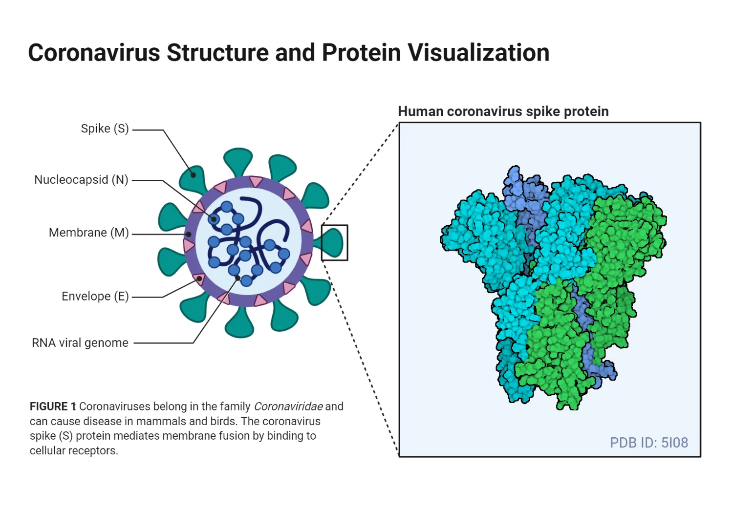
- Major structural proteins:
- Spike (S) protein – a glycoprotein that protrudes from the viral envelope, forming the characteristic crown-like appearance (corona) and mediating *host cell entry.
- Membrane (M) protein – the most abundant structural protein, defines the shape of the viral envelope, and interacts with the nucleocapsid (N) protein to organize assembly.
- Envelope (E) protein – the smallest structural protein, involved in viral assembly, budding, and pathogenesis, and acts as an ion channel (viroporin).
- Nucleocapsid (N) protein – binds to viral RNA to form the helical nucleocapsid, assists in viral RNA packaging, transcription regulation, and modulates host immune response.
- Transmembrane proteins (S and M) help the virus anchor into the lipid envelope and facilitate virus assembly during replication.
Spike Glycoprotein (S protein)
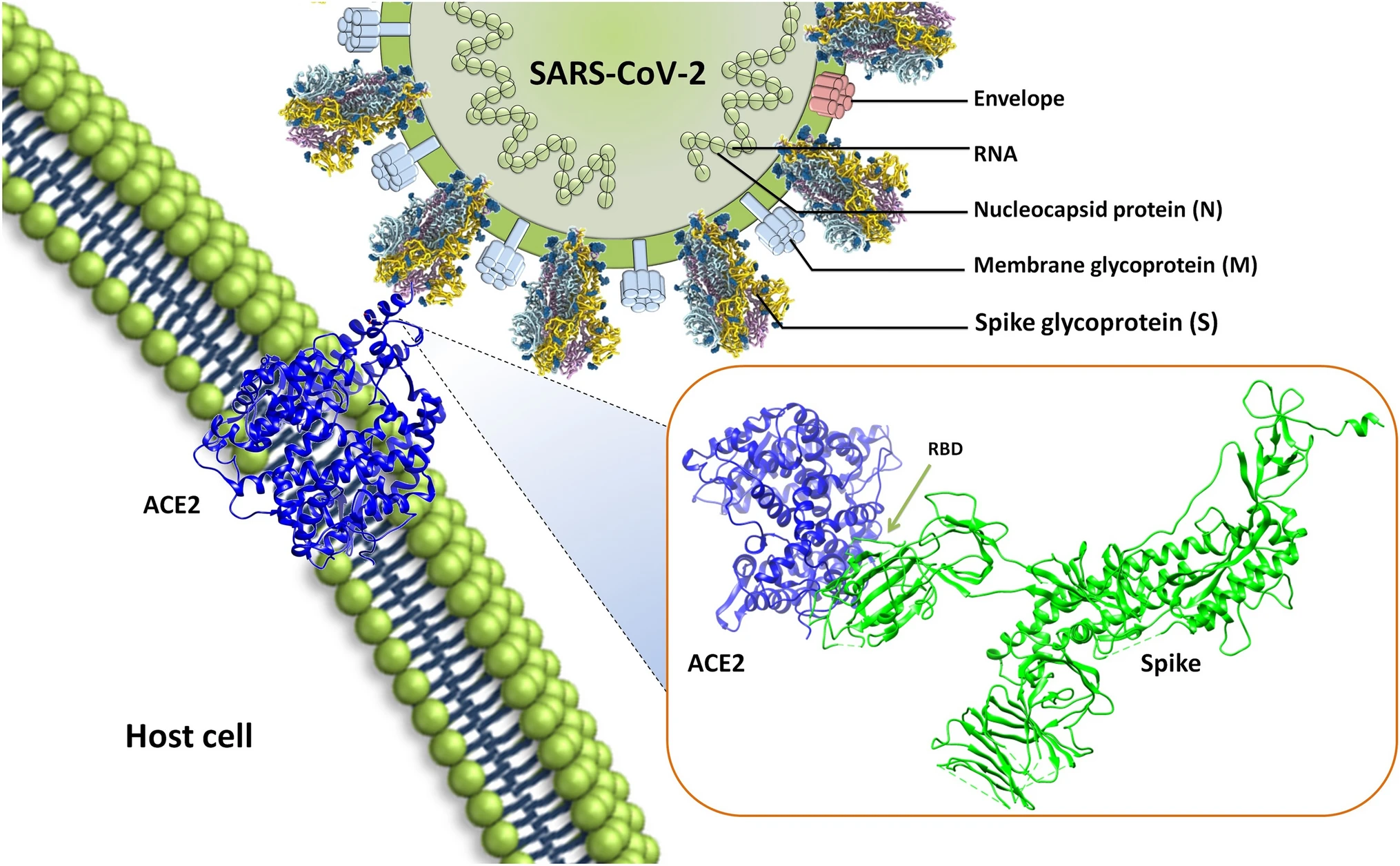
- Comprised of S1 and S2 subunits:
- S1 subunit: contains a signal peptide, N-terminal domain (NTD), and receptor-binding domain (RBD). The RBD binds specifically to ACE2 receptors (Angiotensin-converting enzyme 2) on human respiratory epithelial cells.
- S2 subunit: contains the fusion peptide, heptad repeats (HR1 & HR2), transmembrane domain, and cytoplasmic tail, essential for fusion of viral and host membranes.
- Function: Spike protein mediates host recognition, attachment, and entry, which is critical for viral infectivity.
- Host receptor binding: SARS-CoV-2 binds ACE2 with higher affinity than SARS-CoV, explaining its enhanced transmission.
- After binding, the viral envelope fuses with the host membrane, releasing the viral RNA genome into the cytoplasm.
Genomic Organization
- SARS-CoV-2 has a positive-sense RNA genome of ~30 kb (29,891 nucleotides), encoding 9,860 amino acids, with G+C content ~38%.
- Genome features:
- 12 functional ORFs (open reading frames)
- 9 subgenomic mRNAs
- 5′ and 3′ untranslated regions (UTRs) – regulatory regions for replication and transcription.
- Major ORFs:
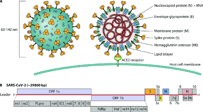
- ORF1a and ORF1b – encode polyproteins pp1a and pp1ab, processed into 16 non-structural proteins (NSPs).
- Remaining ORFs encode structural proteins (S, E, M, N) and accessory proteins, which help the virus evade host immunity.
Key Enzymes and Proteins
- NSP3 – Papain-like protease
- Function: Cleaves viral polyproteins into functional NSPs and suppresses host immune response by deubiquitination.
- NSP5 – Main protease (Mpro)
- Function: Processes viral polyproteins at multiple sites; essential for viral replication.
- NSP12 – RNA-dependent RNA polymerase (RdRp)
- Function: Catalyzes replication of viral RNA, synthesizing complementary negative-sense RNA as a template for progeny genomes.
- NSP13 – Helicase
- Function: Unwinds viral RNA secondary structures during replication and transcription.
- Accessory proteins
- Function: Interfere with host innate immunity, particularly type I interferon response, facilitating immune evasion.
Replication Cycle
- Attachment: Spike protein binds ACE2 receptor → endocytosis or membrane fusion.
- Uncoating: Release of viral RNA into cytoplasm.
- Translation of ORF1a/1b: Polyproteins pp1a/pp1ab are synthesized and cleaved by viral proteases (NSP3 & NSP5) into functional NSPs.
- Replication: RdRp (NSP12) synthesizes negative-sense RNA, used as a template for new positive-sense RNA and subgenomic mRNAs.
- Translation of structural proteins: S, M, E, N are translated and processed through ER-Golgi network.
- Assembly: Nucleocapsid (RNA + N protein) binds M protein at ERGIC (endoplasmic reticulum-Golgi intermediate compartment).
- Budding and Release: Virions bud into ERGIC vesicles → transported via Golgi → released by exocytosis.
Pathogenesis
- Transmission: Respiratory droplets, aerosols, and contact with contaminated surfaces.
- Tropism: Respiratory epithelium (high ACE2 expression).
- Cellular effects: Viral replication leads to ciliostasis (loss of ciliary movement), cell death, and inflammatory cytokine production.
- Severe infection can involve lungs, heart, kidneys, and brain, causing multi-organ failure in critical cases.
Host Interaction
- Immune evasion: Accessory proteins suppress type I interferons, reducing early antiviral response.
- Spike protein: High-affinity binding to ACE2 increases viral entry efficiency.
- Cytokine storm: Overactivation of immune system can lead to tissue damage and ARDS (acute respiratory distress syndrome).
Related posts
More guides connected to this topic

Coronavirus Structure, Genome, Replication, Symptoms, Pathogenesis, and Prevention
Structure of Coronavirus Coronaviruses (enveloped RNA viruses) fall in the virus family Coronaviridae, order Nidovirales. They are enveloped...
Cucumber Mosaic Virus (CMV): Structure, Genome, Symptoms, Disease Cycle and Management
Cucumber Mosaic Virus (CMV): Introduction and Importance Cucumber Mosaic Virus (CMV) (plant-infecting RNA virus) is one of the most economic...
Zoonotic Spillover in Bats: Causes, Consequences, and Future Preparedness
Bats: General Characteristics and Classification Bats are small mammals capable of true sustained flight. They belong to the second-largest...