
On this page
- Nucleic acids (DNA & RNA) are the building blocks of genetic material.
- DNA is the genetic material in most of the organisms.
- RNA is the genetic material in some viruses. RNA mostly functions as messengers.
THE DNA
Structure of Polynucleotide Chain
- Polynucleotides are the polymer of nucleotides.
- DNA & RNA are polynucleotides. A nucleotide has 3 components:
- A nitrogenous base.
- A pentose sugar (ribose in RNA & deoxyribose in DNA).
- A phosphate group.
- Nitrogen bases are 2 types:
- Purines: It includes Adenine (A) and Guanine (G).
- Pyrimidines: It includes Cytosine (C), Thymine (T) & Uracil (U). Thymine (5-methyl Uracil) present only in DNA and Uracil only in RNA.
- A nitrogenous base is linked to the OH of 1' C pentose sugar through an N-glycosidic linkage to form a nucleoside.
- Nucleosides in RNA and DNA:
- RNA: Adenosine → DNA: Deoxyadenosine
- RNA: Guanosine → DNA: Deoxyguanosine
- RNA: Cytidine → DNA: Deoxycytidine
- RNA: Uridine → DNA: Deoxythymidine
- A phosphate group is linked to OH of 5' C of a nucleoside through a phosphoester linkage to form a nucleotide (or deoxynucleotide).
- In RNA, each nucleotide has an additional –OH group at 2' C of the ribose (2'-OH).
- 2 nucleotides are linked through a 3'-5' phosphodiester bond to form a dinucleotide.
- When more nucleotides are linked, it forms a polynucleotide.

Structure of the DNA
- Friedrich Meischer (1869): Identified DNA and named it as 'Nuclein'.
- James Watson & Francis Crick (1953) proposed a double helix model of DNA. It was based on X-ray diffraction data produced by Maurice Wilkins & Rosalind Franklin.


- DNA is made of 2 polynucleotide chains coiled in a right-handed fashion. Its backbone is formed of sugar & phosphates. The bases project inside.
- The 2 chains have anti-parallel polarity, i.e., one chain has the polarity 5'→3' and the other has 3'→5'.
- The bases in 2 strands are paired through H-bonds forming base pairs (bp).
- A=T (2 hydrogen bonds), C≡G (3 hydrogen bonds).
- A purine comes opposite to a pyrimidine. This generates uniform distance between the 2 strands.
- Erwin Chargaff's rule: In DNA, the proportion of A is equal to T and the proportion of G is equal to C.
∴ [A] + [G] = [T] + [C] or [A] + [G] / [T] + [C] = 1.
- Ф 174 (a bacteriophage) has 5386 nucleotides.
- Bacteriophage lambda has 48502 base pairs (bp).
- E. coli has 4.6x10⁶ bp.
- Haploid content of human DNA is 3.3x10⁹ bp.
- Length of DNA = number of base pairs × distance between two adjacent base pairs.
- Number of base pairs in human = 6.6 x 10⁹.
Hence, the length of DNA = 6.6 x 109 x 0.34 x 10-9.
= 2.2 m.
- In E. coli, length of DNA = 1.36 mm (1.36 x 10⁻³ m).
∴ The number of base pairs:
1.36 × 10 − 3 0.34 × 10 − 9
= 4 x 10⁶ bp.
Central Dogma of Molecular Biology
- It is proposed by Francis Crick. It states that the genetic information flows from DNA → RNA → Protein.

Central Dogma of Molecular Biology
- In some viruses, the flow of information is in the reverse direction (from RNA to DNA). It is called reverse transcription.
Packaging of DNA Helix
- In prokaryotes (e.g., E. coli), the DNA is not scattered throughout the cell. DNA is negatively charged. So it is held with some positively charged proteins to form a nucleoid.
- In eukaryotes, there is a set of positively charged, basic proteins called histones.
- Histones are rich in positively charged basic amino acid residues lysines and arginines.
- 8 histones form a histone octamer.
- Negatively charged DNA is wrapped around the histone octamer to give a nucleosome.

- A typical nucleosome contains 200 bp.
- Therefore, total number of nucleosomes in human:

- Nucleosomes constitute the repeating unit to form chromatin. Chromatin is the thread-like stained bodies.
- Nucleosomes in chromatin = 'beads-on-string'.
- Chromatin is packaged → chromatin fibres → coiled and condensed at metaphase stage → chromosomes.
- Higher level packaging of chromatin requires non-histone chromosomal (NHC) proteins.
- Chromatin has 2 forms:
- Euchromatin: Loosely packed and transcriptionally active region of chromatin. It stains light.
- Heterochromatin: Densely packed and inactive region of chromatin. It stains dark.
1. Griffith’s Transforming Principle Experiment (1928)
- Frederick Griffith used mice & Streptococcus pneumoniae.
- Streptococcus pneumoniae has 2 strains:
- Smooth (S) strain (Virulent): Has polysaccharide mucus coat. Causes pneumonia.
- Rough (R) strain (Non-virulent): No mucus coat. Does not cause pneumonia.
- Experiment:
- S-strain → Inject into mice → Mice die.
- R-strain → Inject into mice → Mice live.
- S-strain (Heat killed) → Inject into mice → Mice live.
- S-strain (Heat killed) + R-strain (live) → Inject into mice → Mice die.
- He concluded that some ‘transforming principle’ transferred from heat-killed S-strain to R-strain. It enabled R-strain to synthesize smooth polysaccharide coat and become virulent. This must be due to the transfer of genetic material.
2. Biochemical Characterization of Transforming Principle
- Oswald Avery, Colin MacLeod & Maclyn McCarty worked to determine the biochemical nature of ‘transforming principle’ in Griffith’s experiment.
- They purified biochemicals (proteins, DNA, RNA, etc.) from heat-killed S cells using suitable enzymes.
- They discovered that:
- Digestion of protein and RNA (using Proteases and RNases) did not affect transformation. It means that the transforming substance was not a protein or RNA.
- Digestion of DNA with DNase inhibited transformation. It means that DNA caused transformation of R cells to S cells. It proves that DNA was the transforming principle.
3. Hershey-Chase Experiment (Blender Experiment) - 1952
- Hershey & Chase grew some bacteriophage viruses on a medium containing radioactive phosphorus (P32) and some others on a medium containing radioactive sulphur (S35).
- Viruses grown in P32 got radioactive DNA because only DNA contains phosphorus. Viruses grown in S35 got radioactive protein because protein contains sulphur.
- These preparations were used separately to infect E. coli.
- After infection, the E. coli cells were gently agitated in a blender to remove the virus particles from the bacteria.
- Then the culture was centrifuged to separate lighter virus particles from heavier bacterial cells.
- Bacteria infected with viruses having radioactive DNA were radioactive, i.e., DNA had passed from the virus to bacteria. Bacteria infected with viruses having radioactive proteins were not radioactive, i.e., proteins did not enter the bacteria from the viruses. This proves that DNA is the genetic material.

Properties of Genetic Material (DNA vs RNA)
- A genetic material must have the following properties:
- Ability to generate its replica (Replication).
- Chemical and structural stability.
- Provide the mutations that are required for evolution.
- Ability to express as Mendelian Characters.
- Comparison of DNA and RNA stability and reactivity:
- DNA: Double stranded → RNA: Single stranded.
- DNA: Presence of thymine → RNA: Presence of Uracil.
- DNA: Absence of 2’-OH in sugar → RNA: Presence of 2’-OH in sugar.
- RNA is unstable. So, RNA viruses (e.g., Q.B bacteriophage, Tobacco Mosaic Virus, etc.) mutate and evolve faster.
- DNA strands are complementary. On heating, they separate. In appropriate conditions, they come together. In Griffith’s experiment, some properties of DNA of the heat-killed bacteria were not destroyed. It indicates the stability of DNA.
- For the storage of genetic information, DNA is better due to its stability. But for the transmission of genetic information, RNA is better.
- RNA can directly code for protein synthesis, hence can easily express the characters. DNA is dependent on RNA for protein synthesis.
RNA World
- RNA was the first genetic material.
- It acts as genetic material and catalyst.
- Essential life processes (metabolism, translation, splicing, etc.) evolved around RNA.
- DNA evolved from RNA for stability.
DNA Replication
- Replication is the copying of DNA from parental DNA.
- Watson & Crick proposed the Semi-conservative model of replication. It suggests that the parental DNA strands act as a template for the synthesis of new complementary strands. After replication, each DNA molecule would have one parental and one new strand.
- Matthew Messelson & Franklin Stahl (1958) experimentally proved the Semi-conservative model.
Messelson & Stahl’s Experiment
- They grew E. coli in 15NH4Cl medium (15N = heavy isotope of nitrogen) as the only nitrogen source. As a result, 15N was incorporated into newly synthesised DNA (heavy DNA or 15N DNA).
- Heavy DNA can be distinguished from normal DNA (light DNA or 14N DNA) by centrifugation in a cesium chloride (CsCl) density gradient.
- E. coli cells from the 15N medium were transferred to a 14NH4Cl medium. After one generation (i.e., after 20 minutes), they isolated and centrifuged the DNA. Its density was intermediate (hybrid) between 15N DNA and 14N DNA. This shows that in newly formed DNA, one strand is old (15N type) and one strand is new (14N type). This confirms semi-conservative replication.
- After the second generation (i.e., after 40 minutes), there were equal amounts of hybrid DNA and light DNA.
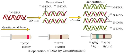
Messelson & Stahl’s Experiment
- Taylor & colleagues (1958) performed similar experiments on Vicia faba (faba beans) using radioactive thymidine to detect the distribution of newly synthesized DNA in the chromosomes. It proved that the DNA in chromosomes also replicates semi-conservatively.
The Machinery and Enzymes for Replication
- DNA replication starts at a point called origin (ori).
- A unit of replication with one origin is called a replicon.
- During replication, the 2 strands unwind and separate by breaking H-bonds in the presence of an enzyme, Helicase.
- Unwinding of the DNA molecule at a point forms a ‘Y’-shaped structure called a replication fork.

Watson-Crick model for semiconservative DNA replication

Replication Fork in DNA Replication
- The separated strands act as templates for the synthesis of new strands.
- DNA replicates in the 5’→3’ direction.
- Deoxyribonucleoside triphosphates (dATP, dGTP, dCTP & dTTP) act as substrates and provide energy for polymerization.
- Firstly, a small RNA primer is synthesized in the presence of an enzyme, primase.
- In the presence of an enzyme, DNA-dependent DNA polymerase, many nucleotides join with one another to the primer strand and form a polynucleotide chain (new strand).
- During replication, one strand is formed as a continuous stretch in the 5’→3’ direction (Continuous synthesis). This strand is called the leading strand.
- The other strand is formed in small stretches (Okazaki fragments) in the 5’→3’ direction (Discontinuous synthesis).
- The Okazaki fragments are then joined together to form a new strand by an enzyme, DNA ligase. This new strand is called the lagging strand.
- If a wrong base is introduced in the new strand, DNA polymerase can do proofreading.
- E. coli completes replication within 18 minutes, i.e., 2000 bp per second.
- In eukaryotes, the replication of DNA takes place at the S-phase of the cell cycle. Failure in cell division after DNA replication results in polyploidy.
Transcription
- It is the process of copying genetic information from one strand of the DNA into RNA.
- Here, adenine pairs with uracil instead of thymine.
- The DNA-dependent RNA polymerase catalyzes the polymerization only in the 5’→3’ direction.
- 3’→5’ acts as the template strand. RNA is built from this.
- 5’→3’ acts as the coding strand. This is copied to RNA.
- 3’-ATGCATGCATGCATGCATGCATGC-5’ template strand.
5’-TACGTACGTACGTACGTACGTACG-3’
- coding strand.
- During transcription, both strands are not copied because:
- The code for proteins is different in both strands. This complicates the translation.
- If 2 RNA molecules are produced simultaneously, this would be complementary to each other. It forms a double-stranded RNA and prevents translation.
Transcription Unit
- It is the segment of DNA between the sites of initiation and termination of transcription. It consists of 3 regions:
- A promoter: Binding site for RNA polymerase. Located towards the 5'-end (upstream).
- Structural gene: The region between promoter and terminator where transcription takes place.
- A terminator: The site where transcription stops. Located towards the 3'-end (downstream).

Transcription unit
- Gene is a functional unit of inheritance. It is the DNA sequence coding for an RNA (mRNA, rRNA, or tRNA).
- Cistron is a segment of DNA coding for a polypeptide during protein synthesis. It is the largest element of a gene.
- Structural gene in a transcription unit is of 2 types:
- Monocistronic structural genes (split genes): It is seen in eukaryotes. Here, coding sequences (exons or expressed sequences) are interrupted by introns (intervening sequences).
- Exons appear in processed mRNA.
- Introns do not appear in processed mRNA.
- Polycistronic structural genes: It is seen in prokaryotes. Here, there are no split genes.
- Monocistronic structural genes (split genes): It is seen in eukaryotes. Here, coding sequences (exons or expressed sequences) are interrupted by introns (intervening sequences).
Transcription in prokaryotes
- In bacteria (prokaryotes), synthesis of all types of RNA is catalyzed by a single RNA polymerase. It has 3 steps:
- Initiation: Here, the enzyme RNA polymerase binds at the promoter site of DNA. This causes the local unwinding of the DNA double helix. An initiation factor (σ factor) present in RNA polymerase initiates the RNA synthesis.
- Elongation: RNA chain is synthesized in the 5’→3’ direction. In this process, activated ribonucleoside triphosphates (ATP, GTP, UTP, & CTP) are added. This is complementary to the base sequence in the DNA template.
- Termination: A termination factor (ρ factor) binds to the RNA polymerase and terminates the transcription.

- In bacteria, transcription and translation can be coupled (translation begins before mRNA is fully transcribed) because:
- mRNA requires no processing to become active.
- Transcription and translation take place in the same compartment (no separation of cytosol and nucleus).
Transcription in eukaryotes
- In eukaryotes, there are 2 additional complexities:
- 1. There are 3 RNA polymerases:
- RNA polymerase I: Transcribes rRNAs (28S, 18S, & 5.8S).
- RNA polymerase II Transcribes the heterogeneous nuclear RNA (hnRNA). It is the precursor of mRNA.
- RNA polymerase III: Transcribes tRNA, 5S rRNA, and snRNAs (small nuclear RNAs).
2. The primary transcripts (hnRNA) contain exons and introns and are non-functional. Hence introns must be removed. For this, it undergoes the following processes:Splicing: From hnRNA, introns are removed (by the spliceosome) and exons are spliced (joined) together.Capping: Here, a nucleotide methyl guanosine triphosphate (cap) is added to the 5’ end of hnRNA.Tailing (Polyadenylation): Here, adenylate residues (200-300) are added at the 3’-end.Now, it is the fully processed mRNA.

Genetic Code
- It is the sequence of nucleotides (nitrogen bases) in mRNA that contains information for protein synthesis (translation).
- The sequence of 3 bases determining a single amino acid is called codon.
- George Gamow suggested that for coding 20 amino acids, the code should be made up of 3 nucleotides. Thus, there are 64 codons (43 = 4 × 4 × 4).
- Har Gobind Khorana developed the chemical method in synthesizing RNA molecules with defined combinations of bases (homopolymers & copolymers).
- Marshall Nirenberg developed a cell-free system for protein synthesis.
- Severo Ochoa (polynucleotide phosphorylase enzyme) is used to polymerize RNA with defined sequences in a template-independent manner.
20 types of amino acids involved in translation
- Alanine (Ala)
- Arginine (Arg)
- Asparagine (Asn)
- Aspartic acid (Asp)
- Cysteine (Cys)
- Glutamine (Gln)
- Glutamic acid (Glu)
- Glycine (Gly)
- Histidine (His)
- Isoleucine (Ile)
- Leucine (Leu)
- Lysine (Lys)
- Methionine (Met)
- Phenylalanine (Phe)
- Proline (Pro)
- Serine (Ser)
- Threonine (Thr)
- Tryptophan (Trp)
- Tyrosine (Tyr)
- Valine (Val)
The codons for various amino acids
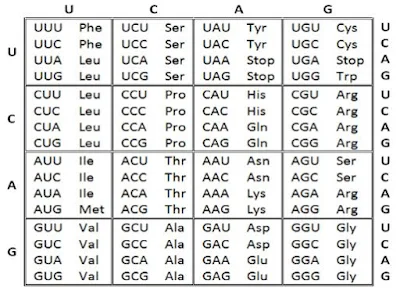
The codons for various amino acids
Salient features of genetic code
- Codon is triplet (three-letter code).
- 61 codons code for amino acids. 3 codons (UAA, UAG & UGA) do not code for any amino acids. They act as stop codons (termination codons or non-sense codons).
- Genetic code is universal. E.g., from bacteria to human, UUU codes for Phenylalanine. Some exceptions are found in mitochondrial codons and in some protozoans.
- No punctuations between adjacent codons (comma-less code). The codon is read in mRNA in a contiguous fashion.
- Genetic code is non-overlapping.
- An amino acid is coded by more than one codon (except AUG for methionine & UGG for tryptophan). Such codons are called degenerate codons.
- Genetic code is unambiguous and specific, i.e., one codon specifies only one amino acid.
- AUG has dual functions. It codes for Methionine and acts as an initiator codon. In eukaryotes, methionine is the first amino acid, and formyl methionine in prokaryotes.
Mutations and Genetic Code
- Relationship between genes & DNA is best understood by mutation studies. Deletions & rearrangements in DNA may cause loss or gain of a gene and thus a function.
- Insertion or deletion of one or two bases changes the reading frame from the point of insertion or deletion. It is called frame-shift insertion or deletion mutations.
- Insertion/deletion of three or its multiple bases inserts or deletes one or multiple codons. The reading frame remains unaltered from that point onwards. Hence, one or multiple amino acids are inserted/deleted.
- It proves that a codon is a triplet and is read contiguously.
Types of RNA
- mRNA (messenger RNA): Provides the template for translation (protein synthesis).
- rRNA (ribosomal RNA): Plays a structural & catalytic role during translation. E.g., 23S rRNA in bacteria acts as a ribozyme.
- tRNA (transfer RNA or sRNA or soluble RNA): Brings amino acids for protein synthesis and reads the genetic code.
- Francis Crick postulated the presence of an adapter molecule that can read the code and link with amino acids.
- tRNA is called an adapter molecule because it has:
- An Anticodon (NODOC) loop that has bases complementary to the codon.
- An amino acid acceptor end to which the amino acid binds.
- Ribosome binding loop.
- Enzyme binding loop.

Structure of tRNA
- For initiation, there is another tRNA called initiator tRNA.
- There are no tRNAs for stop codons.
- Secondary (2-D) structure of tRNA looks like a clover-leaf. The 3-D structure looks like an inverted ‘L’.
Translation (Protein Synthesis)
- It is the process of polymerisation of amino acids to form a polypeptide based on the sequence of codons in mRNA.
- It takes place in ribosomes. Ribosome consists of structural RNAs and about 80 types of proteins.
- Ribosome also acts as a catalyst for the formation of peptide bond between amino acids. E.g., 23S rRNA in bacteria is the enzyme- ribozyme.
- Inactive ribosome exists as two subunits: a large subunit & a small subunit. Large subunit has two sites for subsequent amino acids to bind through peptide bond.
- Formation of peptide bond needs energy from ATP. For this, amino acids are activated (amino acid + ATP) and linked to their cognate tRNA. It is called Charging (aminoacylation) of tRNA.
Steps of Translation
1. Initiation
- In this, small subunit of ribosome binds to mRNA at the start codon (AUG).
- Now large subunit binds to small subunit to form initiation complex.
- Large subunit consists of aminoacyl tRNA binding site (A site) and peptidyl site (P site).
- The initiator tRNA (which carries methionine) binds on P site. Its anticodon (UAC) recognises start codon AUG.

Process of Translation
2. Elongation
- Second aminoacyl tRNA binds to the A site of ribosome. Its anticodon binds to the second codon on the mRNA and a peptide bond is formed between first and second amino acids in presence of peptidyl transferase.
- First amino acid and its tRNA are broken. This tRNA is removed from P site and second tRNA from A site is pulled to P site along with mRNA. This is called translocation.
- These processes are repeated for other codons in mRNA.
- During translation, ribosome moves from codon to codon.
3. Termination
- When a release factor binds to stop codon, the translation terminates.
- The polypeptide and tRNA are released from the ribosomes.
- The ribosome dissociates into large and small subunits.
- A group of ribosomes associated with a single mRNA for translation is called a polyribosome (polysomes).
- An mRNA has additional sequences that are not translated (untranslated regions or UTR). UTRs are present at both 5’-end (before start codon) and 3’-end (after stop codon). They are required for efficient translation process.
Regulation of Gene Expression
- In eukaryotes, gene expression occurs by following levels:
- Transcriptional level (formation of primary transcript).
- Processing level (splicing, capping etc.).
- Transport of mRNA from nucleus to the cytoplasm.
- Translational level (formation of a polypeptide).
- The metabolic, physiological, and environmental conditions regulate gene expression. E.g.:
- In E. coli, the beta-galactosidase enzyme hydrolyses lactose into galactose & glucose. In the absence of lactose, the synthesis of beta-galactosidase stops.
- The development and differentiation of embryo into adult are a result of the expression of several sets of genes.
- If a substrate is added to growth medium of bacteria, a set of genes is switched on to metabolize it. It is called induction.
- When a metabolite (product) is added, the genes to produce it are turned off. This is called repression.
The lac Operon
- An operon is a polycistronic structural gene regulated by a common promoter and regulatory genes. E.g., lac operon, trp operon, ara operon, his operon, val operon etc.
- Lac operon is an operon that controls lactose metabolism in bacteria. It was first elucidated by Francois Jacob & Jacque Monod.
- Lac operon consists of:
- A regulatory or inhibitor (i) gene: Codes for repressor protein.
- 3 structural genes:
- z gene: Codes for beta-galactosidase. It hydrolyses lactose to galactose and glucose.
- y gene: Codes for permease. It increases permeability of the cell to beta-galactosides (lactose).
- a gene: Codes for a transacetylase.
- Genes in the operon function together in the same or related metabolic pathway.
- If there is no lactose (inducer), lac operon remains switched off. The regulator gene synthesizes mRNA to produce repressor protein. This protein binds to the operator region and blocks RNA polymerase movement. So the structural genes are not expressed.
- If lactose or allolactose is provided in the growth medium, it is transported into E. coli cells by the action of permease. Lactose (inducer) binds with repressor protein. So repressor protein cannot bind to operator region. The operator region becomes free and induces the RNA polymerase to bind with promoter. Then transcription starts.
- Regulation of lac operon by repressor is called negative regulation.

In the absence of Inducer

In the presence of Inducer
Human Genome Project (HGP)
- The entire DNA in the haploid set of chromosomes of an organism is called a Genome.
- In Human genome, DNA is packed in 23 chromosomes.
- Human genome contains about 3x10⁹ bp.
- Human Genome Project (1990-2003) was the first mega project for the sequencing of nucleotides and mapping of all the genes in human genome.
- HGP was coordinated by U.S. Department of Energy and the National Institute of Health.
Goals of HGP
- Identify all the estimated genes in human DNA.
- Sequencing of 3 billion chemical base pairs of human DNA.
- Store this information in databases.
- Improve tools for data analysis.
- Transfer related technologies to other sectors.
- Address the ethical, legal, and social issues (ELSI) that may arise from the project.
Methodologies of HGP
- 2 major approaches:
- Expressed Sequence Tags (ESTs): Focused on identifying all the genes that are expressed as RNA.
- Sequence annotation: Sequencing whole set of genome containing all the coding & non-coding sequence and later assigning different regions in the sequence with functions.
- Procedure of sequencing: Isolate DNA from a cell → Convert into random fragments → Clone in a host (bacteria & yeast) using vectors (e.g., BAC & YAC) for amplification → Sequencing of fragments using Automated DNA sequencers (Frederick Sanger method) → Arrange the sequences based on overlapping regions → Alignment of sequences using computer programs.
- BAC = Bacterial Artificial Chromosomes.
- YAC = Yeast Artificial Chromosomes.
- Sanger has also developed a method for sequencing of amino acids in proteins.
- DNA is converted to fragments as there are technical limitations in sequencing very long pieces of DNA.
- HGP was closely associated with Bioinformatics.
- Bioinformatics: Application of computer science and information technology to the field of biology & medicine.
- Of the 24 chromosomes (22 autosomes and X & Y), the last sequenced one is chromosome 1 (May 2006).
- DNA sequencing also has been done in bacteria, yeast, Caenorhabditis elegans (a free-living non-pathogenic nematode), Drosophila, plants (rice & Arabidopsis), etc.
Salient Features of Human Genome
- Human genome contains 3164.7 million nucleotide bases.
- Total number of genes = about 30,000.
- Average gene consists of 3000 bases, but sizes vary. Largest known human gene (dystrophin on X-chromosome) contains 2.4 million bases.
- 99.9% nucleotide bases are the same in all people. Only 0.1% (3x10⁶ bp) difference makes every individual unique.
- Functions of over 50% of discovered genes are unknown.
- Chromosome I has the most genes (2968) and Y has the fewest (231).
- Less than 2% of the genome codes for proteins.
- A very large portion of the human genome is made of repeated (repetitive) sequences. These are stretches of DNA sequences that are repeated many times. They have no direct coding functions. They shed light on chromosome structure, dynamics, and evolution.
- About 1.4 million locations have single-base DNA differences. They are called SNPs (Single nucleotide polymorphism or ‘snips’). This helps to find chromosomal locations for disease-associated sequences and tracing human history.
DNA Fingerprinting (DNA Profiling)
- It is the technique to identify the similarities and differences of the DNA fragments of 2 individuals.
- It is developed by Alec Jeffreys (1985).
Basis of DNA Fingerprinting
- DNA carries some non-coding repetitive sequences.
- Repetitive DNA can be separated from bulk genomic DNA as different peaks during density gradient centrifugation.
- The bulk DNA forms a major peak and the small peaks are called satellite DNA.
- Satellite DNA is classified as micro-satellites, mini-satellites etc. based on base composition (A:T rich or G:C rich), length of segment, and number of repetitive units.
- A DNA sequence which is tandemly repeated in many copy numbers is called variable number tandem repeats (VNTR). It belongs to mini-satellite DNA.
- In a person, copy number varies in each chromosome.
- The two alleles (paternal and maternal) of a chromosome also contain different copy numbers of VNTR.
- VNTR is specific from person to person.
- The size of VNTR varies from 0.1 to 20 kb.
- Any difference in the nucleotide sequence (inheritable mutation) observed in a population is called DNA polymorphism (variation at genetic level).
- Polymorphism is higher in non-coding DNA sequence because mutations in these sequences may not affect an individual’s reproductive ability. These mutations accumulate generation to generation causing polymorphism.
- Polymorphisms have a great role in evolution & speciation.
Steps of DNA Fingerprinting (Southern Blotting Technique)
- Isolation of DNA (from any cells or blood stains, semen stains, saliva, hair roots, bone, skin, etc.).
- Digestion of DNA by restriction endonucleases.
- Separation of DNA fragments by gel electrophoresis.
- Transferring (blotting) DNA fragments to synthetic membranes such as nitrocellulose or nylon.
- Hybridization using radioactive labelled VNTR probe.
- Detection of hybridized DNA by autoradiography.
- The autoradiogram gives an image in the form of dark & light bands. It is called DNA fingerprint.

- DNA fingerprint differs in everyone except in monozygotic (identical) twins.
- The sensitivity of the technique can be increased by the use of polymerase chain reaction (PCR). Therefore, DNA from a single cell is enough for DNA fingerprinting.
Application of DNA Fingerprinting
- Forensic tool to solve paternity, rape, murder, etc.
- For the diagnosis of genetic diseases.
- To determine the phylogenetic status of animals.
- To determine population and genetic diversities.
Related posts
More guides connected to this topic
Biodiversity and Conservation Biology Notes - Levels, Patterns, Loss, Conservation, Hotspots and Protected Areas
Learn about biodiversity levels, patterns, loss, conservation strategies, hotspots and protected areas, with examples from India.
Microbes in Human Welfare Biology Notes - Household, Industrial, Sewage Treatment, Biogas, Biocontrol and Biofertilizers
Explore how microbes are used in household products, food fermentation, industrial processes, sewage treatment, biogas, biocontrol and biofe...
Human Health and Diseases Biology Notes - Common Diseases, Immunity, AIDS, Cancer, Drugs and Adolescence
Overview of common diseases, immune system basics, AIDS, cancer, drug effects and health considerations for adolescents.

Discussion
Comments
Please log in to join the discussion.
Login to commentNo comments yet. Be the first to start the discussion.