
On this page
A group of cells having the same origin, structure, and function are called tissues.
Animal tissues are classified into the following types:
- Epithelial
- Connective
- Muscular
- Neural
I. Epithelial Tissue (Epithelium)
- It has a free surface that faces body fluid or the outside environment.
- Covers or lines the body or body parts.
- Compactly packed cells with little intercellular matrix.
- Epithelial tissues are 2 types: Simple and Compound.
A. Simple Epithelium
- It is composed of a single layer of cells. It lines body cavities, ducts and tubes.
- Based on structural modification of cells, simple epithelium is 3 types:
Squamous Epithelium
- Thin layer of flattened cells with irregular boundaries.
- Found in the walls of blood vessels and lung alveoli.
- Functions: Form a diffusion boundary.
Cuboidal (cubical) Epithelium
- Composed of cube-like cells.
- Found in ducts of glands and tubular parts of nephrons.
- Functions: Secretion and absorption.
- The epithelium of proximal convoluted tubule (PCT) of nephron in the kidney has microvilli.
Columnar Epithelium
- Composed of tall and slender cells.
- Their nuclei are located at the base.
- Free surface may have microvilli.
- Found in the lining of the stomach and intestine.
- Functions: Secretion and absorption.

Modifications of Columnar or Cuboidal Cells:
- Ciliated Epithelium:
- Cells bearing cilia on their free surface.
- Present in the inner surface of hollow organs like bronchioles and fallopian tubes.
- Functions: To move particles or mucus in a specific direction over the epithelium.
- Glandular Epithelium: For secretion. They are of two types:
- Unicellular: Consists of isolated glandular cells. E.g., Goblet cells of the alimentary canal.
- Multicellular: Contains a cluster of cells. E.g., salivary glands.
- Based on the mode of secretion, glands 2 types:
- Exocrine Glands: Secretions are released through ducts (tubes). They secrete mucus, saliva, earwax, oil, milk, digestive enzymes, etc.
- Endocrine Glands: Ductless. They produce hormones.

B. Compound Epithelium
- Made up of more than one layer (multi-layered) of cells.
- Limited role in secretion and absorption.
- They cover the dry surface of the skin, the moist surface of the buccal cavity, pharynx, inner lining of ducts of salivary glands, and pancreatic ducts.
- Function: Protect against chemical and mechanical stresses.

Cell Junctions
- The junctions that provide structural and functional links between adjacent cells.
- Found in epithelium and other tissues.
- They are three types:
- Tight Junctions: Help to stop substances from leaking across a tissue.
- Adhering Junctions: Perform cementing to keep neighboring cells together.
- Gap Junctions: Facilitate communication between adjoining cells by connecting the cytoplasm for rapid transfer of ions, small molecules, and sometimes big molecules.
II. Connective Tissue
- It links and supports other tissues/organs.
- They are most abundant in complex animals.
- All connective tissues except blood have fibroblast cells. They secrete structural fibrous proteins called collagen & elastin. They give strength, elasticity & flexibility to tissue.
- The cells also secrete modified polysaccharides (matrix), which accumulate between cells and fibres.
- Types of connective tissues: Loose, Dense & Specialised.
1. Loose Connective Tissues
In this, cells (fibroblasts, macrophages, mast cells etc.) and fibres are loosely arranged in a semi-fluid matrix.
It is of 2 types:
Areolar tissue:
- Present beneath the skin.
- It serves as a support framework for epithelium.

Adipose tissue:
- Seen mainly under skin.
- Its cells (adipocytes) store fats.
- Excess nutrients which are converted into fats are stored in this tissue.

2. Dense Connective Tissues
In this, fibres and fibroblasts are compactly packed.
It is of 2 types:
Dense regular connective tissues:
- Show regular pattern of fibres.
- Collagen fibres are present in rows between many parallel bundles of fibres.
- E.g. tendons & ligaments.
- Tendons: Attach muscles to bones.
- Ligaments: Attach one bone to another.

Dense irregular connective tissues:
- Irregular pattern of fibres.
- Fibroblasts & fibres (mostly collagen) are oriented differently.
- This tissue is present in skin.

3. Specialized Connective Tissues
Cartilage:
- In this, intercellular material (matrix) is solid and pliable (due to chondroitin salts) and resists compression.
- Cartilage cells (chondrocytes) are enclosed in small cavities within the matrix secreted by them.
- Most of the cartilages in vertebrate embryos are replaced by bones in adults.
- Cartilage is present in the tip of nose, outer ear, joints in the vertebral column, limbs and hands in adults.

Bone:
- It has hard and non-pliable matrix rich in calcium salts and collagen fibres which give bone its strength.
- Bone cells (osteocytes) are seen in spaces called lacunae.
- Functions:
- It provides structural frame to the body.
- Support and protect softer tissues and organs.
- Limb bones serve weight-bearing functions.
- Take part in locomotion and movements.
- Blood cells are produced in bone marrow.

Blood:
- A fluid connective tissue containing plasma, red blood cells (RBC), white blood cells (WBC) and platelets.
- Helps in the circulation of various substances.
III. Muscle Tissue
- The tissues made of many muscle fibres (muscle cells).
- Muscle fibres are composed of numerous fine myofibrils.
- Muscle fibres can contract (shorten) and relax (lengthen).
- Muscles take part in locomotion and movements.
- Muscles are 3 types: skeletal, smooth and cardiac.

1. Skeletal (striated or voluntary) muscle
- They are attached to bones. E.g. Biceps.
- Striations are present in muscle fibres.
- Muscle fibres are bundled together in a parallel fashion.
- A sheath of tough connective tissue encloses several bundles of muscle fibres.
2. Smooth (non-striated or visceral) muscle
- Involuntary and fusiform (Fibres taper at both ends).
- No striations.
- Cell junctions hold them together and they are bundled together in a connective tissue sheath.
- They are seen in the wall of internal organs such as the blood vessels, stomach and intestine.
3. Cardiac muscle
- Involuntary muscle seen only in the heart.
- Cell junctions fuse the plasma membranes of cardiac muscle cells and make them stick together.
- Communication (gap) junctions (intercalated discs) at some fusion points allow cells to contract as a unit, i.e., when a cell receives signal to contract, other cells also contract.
IV. Neural Tissue
- Made up of neurons (unit of neural system).
- Responsible for control and coordination of the body.
- Neurons are excitable cells. They carry impulses.
- Neurons are protected and supported by neuroglial cells.
- Neuroglia make up more than half the volume of neural tissue.
Organ and Organ System
- Cells → tissues → organs → organ systems.
- This organization is essential for better coordinated activities of cells.
- An organ is made of one or more type of tissues. E.g. Heart has epithelial, connective, muscular & neural tissues.
- Morphology: Study of external features of organisms.
- Anatomy: Study of morphology of internal organs.
- Cockroach (Periplaneta americana) are nocturnal, omnivores and live in damp places.
- Colour: Brown or black. Bright yellow, red & green coloured cockroaches are also seen in tropical regions.
- Size: ¼ inches to 3 inches (0.6-7.6 cm).
Morphology of Cockroach
- The adults are about 34-53 mm long.
- Body is covered by a hard brown chitinous exoskeleton.
- In each segment, exoskeleton has hardened plates called sclerites (dorsal tergites & ventral sternites). They are joined to each other by a thin and flexible articular membrane (arthrodial membrane).
- The body has 3 regions – head, thorax and abdomen.

Head
- Triangular head is formed by 6 fused segments.
- It shows great mobility in all directions due to flexible neck.
- Head bears a pair of thread-like antennae, a pair of compound eyes and biting & chewing type mouth parts.

Mouthparts
- A labrum (upper lip), 2 mandibles, 2 maxillae, hypopharynx (tongue) & a labium (lower lip).

Thorax
- It has 3 parts: prothorax, mesothorax & metathorax.
- The head is connected to thorax by a neck (short extension of the prothorax).
- Each thoracic segment bears a pair of walking legs.
- 2 pairs of wings: Forewings (2) and Hind wings (2).
- Forewings (mesothoracic) or tegmina: Opaque, dark and leathery and cover the hind wings when at rest.
- Hind wings (metathoracic): Transparent, membranous and are used in flight.
Abdomen
- It consists of 10 segments.
- In females, 7th (boat shaped), 8th & 9th sterna form a brood (genital) pouch. It contains female gonopore, spermathecal pores & collateral glands.
- In males, genital pouch lies at the hind end of abdomen bounded dorsally by 9th & 10th terga and ventrally by the 9th sternum. It contains dorsal anus, ventral male genital pore (gonopore) and gonapophysis.
- In both sexes, 10th segment bears a pair of jointed anal cerci. Males bear a pair of short, threadlike anal styles.
Differences between Male & Female Cockroaches
| Male | Female |
|---|---|
| Larger size | Smaller |
| Wings extend beyond the tip of the abdomen. | Wings do not extend beyond the tip of abdomen. |
| Narrow abdomen | Broad abdomen |
| Anal styles present | Absent |
| Brood pouch absent | Present |
Digestive System
Alimentary canal has 3 parts: foregut, mid gut & hindgut.
- Foregut: It is lined by cuticle. It includes Mouth → pharynx → oesophagus → crop (to store food) → gizzard (proventriculus).
Gizzard helps in grinding the food. It has an outer layer of thick circular muscles and thick inner cuticle forming 6 chitinous plates (teeth).
- Mid gut (Mesenteron): It is not lined by cuticle. 6-8 tubules (hepatic or gastric caecae) are seen at the junction of foregut & mid gut. They secrete digestive juice.
At the junction of mid gut & hindgut, there are 100-150 yellow coloured thin filamentous Malpighian tubules.
- Hindgut: It is broader than mid gut and lined internally by cuticle. Hindgut includes ileum, colon & rectum. Rectum opens out through anus.

Circulatory System
- Blood vascular system: open type.
- Blood vessels are poorly developed and open into space (haemocoel).
- Visceral organs located in the haemocoel are bathed in blood (haemolymph).
- Haemolymph= colourless plasma + haemocytes.
- Heart consists of elongated muscular tube lying along mid dorsal line of thorax and abdomen.
- It has funnel-shaped chambers with ostia on either side.
- Blood from sinuses enter heart through ostia and is pumped anteriorly to sinuses again.

Respiratory System
- It consists of a network of trachea that open through 10 pairs of small holes called spiracles present on the lateral side of the body.
- The thin branches of tracheal tubes are called tracheoles. They carry oxygen from the air to all parts.
- The opening of the spiracles is regulated by sphincters.
- Gas exchange takes place at the tracheoles by diffusion.
Excretory System
- Uricotelic. Excretory organ is Malpighian tubules.
- Each tubule is lined by glandular and ciliated cells. They absorb nitrogenous wastes and convert them into uric acid which is excreted out through the hindgut.
- Fat body, nephrocytes & urecose glands also help in excretion.
Nervous System
- It consists of segmentally arranged ganglia joined by paired longitudinal connectives on the ventral side.
- 3 ganglia lie in the thorax and 6 in the abdomen.
- The head holds only a bit of nervous system. Remaining part is situated along the ventral part of the body. So, if the head of cockroach is cut off, it will still live for one week.
- The supra-oesophageal ganglion (brain) supplies nerves to antennae and compound eyes.
- Sense organs: Antennae, eyes, maxillary palps, labial palps, anal cerci etc.
- Sensory receptors of antennae monitor the environment.
- Each compound eye consists of about 2000 hexagonal ommatidia. Using these, a cockroach can receive several images of an object. This is called mosaic vision. It has more sensitivity but less resolution, being common during night (hence called nocturnal vision).
Reproductive System
- Cockroaches are dioecious.
Male Reproductive System
- It consists of a pair of testes, seminal vesicles, accessory glands & external genitalia.
- Testes: Lie laterally in the 4th -6th abdominal segments.
Each testis → a thin vas deferens → seminal vesicle → ejaculatory duct → male gonopore.
- Seminal vesicles: To store sperms. Sperms are glued together to form bundles called spermatophores. They are discharged during copulation.
- Accessory glands: Include a mushroom gland (in 6th-7th abdominal segments) and phallic gland. Their secretions nourish the sperms.
- External genitalia (gonapophysis or phallomeres): Chitinous asymmetrical structures, surrounding the male gonopore.

Female Reproductive System
- It consists of 2 large ovaries, oviducts, spermatheca, genital chamber, Colleterial glands etc.
- Ovaries lie laterally in the 2nd – 6th abdominal segments. Each ovary is formed of 8 ovarian tubules (ovarioles), containing a chain of developing ova.
- Oviducts of each ovary unite into a single median oviduct (vagina) which opens into the genital chamber.
- A pair of spermatheca is present in the 6th segment which opens into the genital chamber.
- Sperms are transferred through spermatophores. Their fertilised eggs are encased in oothecae.
- Ootheca is dark reddish to blackish brown capsule, 8 mm long. Females lay 9-10 oothecae, each contain 14-16 eggs.
- Development of P. americana is paurometabolous (development through nymphal stage).
- Nymphs look like adults. They moult 13 times to reach the adult form.
- The next to last nymphal stage has wing pads. Only adult cockroaches have wings.

Economic Importance of Cockroach
- They are pests because they destroy food and contaminate it with their smelly excreta.
- They also transmit bacterial diseases like cholera, typhoid, tuberculosis etc.
- Earthworm is a reddish-brown terrestrial invertebrate that inhabits the upper layer of moist soil.
- During day time, they live in burrows made by boring and swallowing the soil.
- Common Indian earthworms: Pheretima and Lumbricus.
Morphology of Earthworm
- Earthworms have long segmented cylindrical body.
- Number of segments (metameres): about 100-120.
- Dorsal surface has a dark median mid dorsal line (dorsal blood vessel) along the longitudinal axis of the body.
- First segment (peristomium or buccal segment) bears the mouth. A lobe called prostomium covers the mouth.
- Prostomium is sensory in function and is used to force open cracks in the soil into which the earthworm may crawl.
- In a mature worm, segments 14-16 are covered by a dark band of glandular tissue called clitellum.
- Body has 3 regions: preclitellar, clitellar & postclitellar.
- 4 pairs of spermathecal apertures are found on ventro-lateral sides of intersegmental grooves (5th -9th segments).
- A single female genital pore is present in the mid-ventral line of 14th segment.
- A pair of male genital pores is present on the ventro-lateral sides of the 18th segment.
- Many minute nephridiopores open on the body surface.
- All segments except the first, last, and clitellum bear S-shaped setae, embedded in the epidermal pits. Setae can be extended or retracted. Their function is locomotion.

Body Wall
- It is composed of:
- Outermost thin non-cellular cuticle.
- Epidermis: Made up of a single layer of columnar epithelial cells which contain secretory gland cells.
- Two muscle layers (circular and longitudinal).
- An innermost coelomic epithelium.
Digestive System
- The straight alimentary canal extends from first to last segment of the body. It has:
Mouth → buccal cavity (1-3 segments) → muscular pharynx (4th segment) → oesophagus (5-7 segments) → muscular gizzard (8-9 segments) → stomach (9-14 segments) → Intestine (15th segment to last) → anus.
- Gizzard helps to grind soil particles, decaying leaves, etc.
- Calciferous glands, present in the stomach, neutralise the humic acid present in humus.
- A pair of short and conical intestinal caecae project from the intestine on the 26th segment.
- The intestinal part between 26-35 segments has an internal median fold of dorsal wall called typhlosole. It increases area of absorption.
- The organic rich soil is digested in the digestive tract by digestive enzymes. Digested nutrients are absorbed through intestinal membranes. Their faecal deposits are known as worm castings.

Circulatory System (Blood Vascular System)
- Circulatory system is closed type (blood flows through heart and blood vessels).
- Consists of blood vessels, capillaries and heart.
- Contractions keep blood circulating in one direction.
- Blood glands are present on the 4th, 5th & 6th segments. They produce phagocytic blood cells and haemoglobin which is dissolved in blood plasma.

Respiratory System
- No specialized system.
- Gas exchange occurs through moist body surface into the blood stream.
Excretory System
- Excretory organs are segmentally arranged tubules called nephridia. They are 3 types:
- Septal nephridia: Found on both sides of intersegmental septa (segment 15 to last) that open into intestine.
- Integumentary nephridia: Attached to lining of body wall (segment 3 to last). They open on body surface.
- Pharyngeal nephridia: Present in the 4th, 5th & 6th segments in the form of paired tufts.
- Funnel-shaped part of nephridium collects excess fluid from coelom. The funnel connects with a tubular part of nephridium which delivers the wastes into digestive tube.

Nervous System
- Includes segmentally arranged ganglia on the ventral paired and fused nerve cord.
- The nerve cord in the anterior region (3rd & 4th segments) divides and encircles the pharynx and joins the cerebral ganglia dorsally to form a nerve ring.
- The nerve ring with cerebral ganglia represents the brain.
- Sensory system: Includes:
- Light and touch sensitive receptor cells. No eyes.
- Chemoreceptors (taste receptors): React to chemical stimuli.
Reproductive System
- Earthworm is hermaphrodite.
Male reproductive organs:
- Testes: 2 pairs. Enclosed in testis sacs in 10th & 11th segments. The sperms from testes shed into testis sacs. From where, they enter seminal vesicles for maturation. Mature sperms move back into testis sacs and enter spermiducal funnels which are connected to vasa deferentia (spermatic ducts). The vasa deferentia run up to 18th segment where they join the prostatic duct.
The common prostate and spermatic duct open to the exterior by a pair of male genital pores on the ventro-lateral side of the 18th segment.
- Accessory glands: 2 pairs. Found in the 17th & 19th segments (one pair in each segment).
Female reproductive organs:
- Spermathecae: 4 pairs. Located in 6th-9th segments (one pair in each segment). They receive and store spermatozoa during copulation.
- Ovaries: Paired. Attached at the inter-segmental septum of the 12th and 13th segments.
- Ovarian funnels: Present beneath the ovaries which continue into oviduct, join together and open on ventral side as single median female genital pore on 14th segment.
- During mating, two worms exchange sperms each other. They mate juxtaposing opposite gonadal openings exchanging spermatophores (packets of sperms).
- Mature sperm, ova and nutritive fluid are deposited in cocoons produced by gland cells of clitellum. Cocoon with fertilized ova slips off and deposit in the soil. After 3 weeks, cocoon produces 2 to 20 baby worms (no larva).

Economic Importance
- Earthworms are known as ‘friends of farmers’ because they make burrows in the soil and make it porous which helps in respiration and penetration of the plant roots. This process of increasing fertility of soil is called vermicomposting.
- They are used as bait in game fishing.
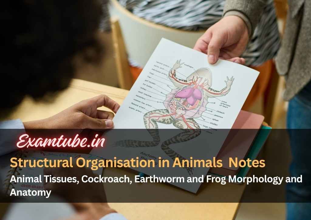
- Rana tigrina is the most common species in India.
- They are poikilotherms (cold blooded).
- They can change colour to hide from their enemies (camouflage). This protective coloration is called mimicry.
- During summer and winter, they undergo aestivation (summer sleep) and hibernation (winter sleep) respectively to protect them from extreme heat and cold.
Morphology of Frog
- Body is divisible into head and trunk. Neck and tail absent.
- Skin is moist, smooth, and slippery due to the mucus.
- Colour of dorsal side is olive green with dark irregular spots, and ventral side is pale yellow.
- The frog never drinks water but absorbs it through the skin.
- A mouth, paired nostrils, and bulged eyes (covered by nictitating membrane) are present.
- On either side of eyes have a membranous tympanum (ear).
- The forelimbs (4 digits) and hind limbs (5 digits) help in swimming, walking, leaping, and burrowing. The hind limbs are larger and muscular than forelimbs.
- Feet have webbed digits that help in swimming.
- Frogs exhibit sexual dimorphism. Male frogs have sound-producing vocal sac and also a copulatory (nuptial) pad on the first digit of forelimbs, which are absent in female frogs.
Anatomy of Frog
Digestive System
- Consists of alimentary canal and digestive glands.
- The alimentary canal is short because frogs are carnivores, hence the length of intestine is reduced.
- Mouth → buccal cavity → pharynx → oesophagus → stomach → intestine → rectum → cloaca.
- Liver secretes bile that is stored in gall bladder. Pancreas produces pancreatic juice containing digestive enzymes.
- Food is captured by the bilobed tongue.
- Digestion: Gastric juice and HCl secreted from gastric wall digest the food. Partially digested food (chyme) is passed from stomach to the duodenum.
- Duodenum receives bile and pancreatic juices through a common bile duct.
- Bile emulsifies fat. Pancreatic juice digests carbohydrates and proteins. Digestion completes in the intestine.
- Finger-like villi and microvilli in intestine absorb digested food. The undigested solid waste moves into the rectum and passes out through cloaca.

Respiratory System
- Skin acts as aquatic respiratory organ (cutaneous respiration). Dissolved oxygen in the water is exchanged through the skin by diffusion. During aestivation and hibernation, respiration takes place through skin.
- On land, the buccal cavity, skin, and lungs (pulmonary respiration) act as the respiratory organs.
- The lungs are a pair of elongated, pink coloured sac-like structures present in the thorax. Air enters through the nostrils into the buccal cavity and then to lungs.
Circulatory System
- Closed type. Includes Blood vascular system (heart, blood vessels & blood) and lymphatic system (lymph, lymph channels & lymph nodes).
- Heart is 3-chambered, (two atria and one ventricle) and is covered by a membrane called pericardium.
- A triangular structure called sinus venosus joins the right atrium. It receives blood through major veins (vena cava).
- The ventricle opens into a saclike conus arteriosus on the ventral side of the heart.
- The blood pumped from the muscular heart is carried to all parts of the body by the arteries (arterial system).
- The veins collect blood from different parts of body to the heart and form the venous system.
- Hepatic portal system (venous connection between liver and intestine) and renal portal system (between kidney and lower parts of the body) are present in frogs.
- Blood contains plasma and cells (RBC, WBC & platelets). RBCs are nucleated and contain haemoglobin.
- Blood transports nutrients, gases, and water to tissues.
Excretory System
- Includes kidneys (2), ureters (2), cloaca & urinary bladder.
- Kidneys are dark red and bean-shaped. Found posteriorly in the body cavity on both sides of vertebral column. Each kidney is formed of uriniferous tubules (nephrons).
- 2 ureters emerge from the kidneys. In male frogs, the ureters act as urinogenital duct which opens into cloaca.
- In females, ureters & oviduct open separately in cloaca.
- The thin-walled urinary bladder is present ventral to the rectum which also opens in the cloaca.
- The frog is a ureotelic animal (excretes urea). Nitrogenous wastes are carried by blood into the kidney where it is separated and excreted.
Control and Coordination
Endocrine System
- The endocrine glands secrete hormones.
- Endocrine glands: pituitary, thyroid, parathyroid, thymus, pineal body, pancreatic islets, adrenals & gonads.
Nervous System
- It includes:
- Central nervous system (brain & spinal cord),
- Peripheral nervous system (cranial & spinal nerves),
- Autonomic nervous system (sympathetic & parasympathetic).
- There are 10 pairs of cranial nerves arising from brain.
- Brain is enclosed in a bony brain box (cranium).
- The brain is divided into:
- Fore-brain: Includes olfactory lobes, paired cerebral hemispheres, and unpaired diencephalon.
- Mid-brain: Includes a pair of optic lobes.
- Hind-brain: Includes cerebellum & medulla oblongata.
- Medulla oblongata passes out through the foramen magnum and continues into spinal cord, which is enclosed in the vertebral column.
- Sense organs include organs of:
- Sensory papillae: For touch,
- Taste buds: For taste,
- Nasal epithelium: For smell,
- Simple eyes: For vision. Paired and situated in orbit,
- Tympanum with internal ears: For hearing and balancing (equilibrium).
Reproductive System
- Male reproductive organs consist of a pair of yellowish ovoid testes, which are found adhered to the upper part of kidneys by a double fold of peritoneum (mesorchium).
- Vasa efferentia (10-12 in number) arise from testes. They enter the kidneys on their side and open into Bidder’s canal. It communicates with urinogenital duct that comes out of the kidneys and opens into cloaca.
- The cloaca is a small, median chamber that is used to pass faecal matter, urine, and sperms to the exterior.

- The female reproductive organs include a pair of ovaries. The ovaries are situated near kidneys, and there is no functional connection with kidneys.
- A pair of oviduct arising from the ovaries opens into the cloaca separately.
- A mature female can lay 2500 to 3000 ova at a time.
- Fertilisation is external and takes place in water.
- Development involves a larval stage called tadpole.
- Tadpole undergoes metamorphosis to form the adult.

Economic Importance
- Frogs are beneficial for mankind because they eat insects and protect the crop.
- Maintain ecological balance by serving as an important link of food chain and food web in the ecosystem.
- In some countries, the muscular legs of frog are used as food by man.
Related posts
More guides connected to this topic
Chemical Coordination and Integration Biology Notes - Endocrine System, Hormones, Glands and Mechanism of Hormone Action
Overview of endocrine glands, hormone types, hypothalamus‑pituitary link and mechanisms of hormone action.
Neural Control and Coordination Biology Notes - Neuron Structure, Nerve Impulse, Synapse, Brain, Reflex and Sensory Organs
Explains neuron anatomy, types, axon variations, nerve impulse transmission, synapses, brain and reflex pathways.
Locomotion and Movement Biology Notes - Muscle Types, Sarcomere Structure, Sliding Filament Theory, Skeletal System and Joints
Covers voluntary locomotion, muscle classifications, sarcomere structure, sliding filament theory and skeletal joint basics.

Discussion
Comments
Please log in to join the discussion.
Login to commentNo comments yet. Be the first to start the discussion.