
On this page
Excretion is the elimination of metabolic wastes like ammonia, urea, uric acid etc. from the tissues.
Types of Excretion
1. Ammonotelism
- Process of excretion of Ammonia (NH3).
- Ammonotelic animals: Aquatic invertebrates, aquatic insects, bony fishes, aquatic amphibians etc.
- NH3 is highly toxic. So, excretion needs excess of water.
- NH3 is readily soluble in water and is excreted by diffusion through body surface or gill surfaces (in fishes) as ammonium ions.
- Kidneys do not play any significant role in its removal.
2. Ureotelism
- Process of excretion of urea.
- Ureotelic animals: Cartilaginous fishes, terrestrial & semi-aquatic amphibians (frogs, toads etc.), aquatic & semi-aquatic reptiles (alligators, turtles), mammals etc.
- In liver, NH3 is converted into less toxic urea. So, it needs only moderate quantity of water for excretion.
- Some amount of urea may be retained in the kidney matrix of some animals to maintain a desired osmolarity.
3. Uricotelism
- Process of excretion of uric acid.
- It is water insoluble & less toxic. So, water is not needed for excretion.
- Uricotelic animals: Insects, some land crustaceans, land snails, terrestrial reptiles & birds.
- Ureotelism & uricotelism are needed for water conservation.
Some Excretory Organs in Animals
- Protonephridia (flame cells): In Flatworms, rotifers, some annelids & cephalochordate (Amphioxus). Protonephridia are primarily for osmoregulation.
- Nephridia: In Annelids. Help in the removal of nitrogenous wastes and osmoregulation.
- Malpighian tubules: In Insects. Help in the removal of nitrogenous wastes and osmoregulation.
- Antennal or green glands: In Crustaceans (prawn etc.)
- Kidneys: In higher animals.
Human Excretory System
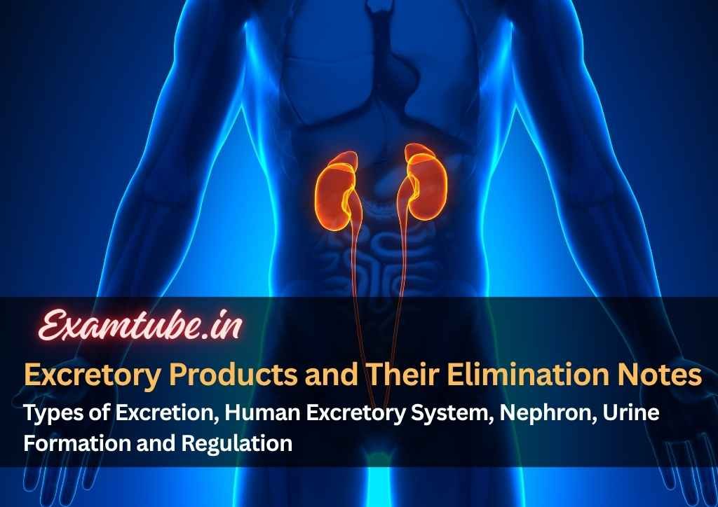
It includes kidneys, ureters, urinary bladder & urethra.

Structure of Kidney
- Reddish brown, bean-shaped structures situated between the levels of last thoracic & 3rd lumbar vertebra.
- Length: 10-12 cm, width: 5-7 cm, thickness: 2-3 cm. Average weight: 120-170 gm.
- It is enclosed in a tough, 3-layered fibrous renal capsule.
- On the concave side of kidney, there is an opening (hilum or hilus) through which blood vessels, nerves, lymphatic ducts and ureter enter the kidney.
- Hilum leads to funnel shaped cavity called renal pelvis with projections called calyces.
- A kidney has outer cortex & inner medulla.

- Medulla has few conical projections called medullary pyramids (renal pyramids) projecting into the calyces.
- Cortex extends in between the medullary pyramids as renal columns (Columns of Bertini).
- Each kidney has nearly one million tubular nephrons.
Nephron

- Nephrons are the structural & functional units of kidney.
- Each nephron has 2 parts: Glomerulus & Renal tubule.
- Glomerulus: A tuft of capillaries formed by afferent arteriole (a fine branch of renal artery). Blood from glomerulus is carried away by efferent arteriole.
- Renal tubule: It begins with a double walled cup-like Bowman’s capsule, which encloses the glomerulus.
Glomerulus + Bowman’s capsule = Malpighian body
- The tubule continues with proximal convoluted tubule (PCT), Henle’s loop & distal convoluted tubule (DCT).
- Henle’s loop is hairpin-shaped. It has descending and ascending limbs.
- The DCTs of many nephrons open into a collecting duct. Collecting duct extends from cortex to inner parts of medulla. They converge and open into the renal pelvis through medullary pyramids in the calyces.
- lip>
Malpighian body (Renal corpuscle),
PCT
- and
DCT
- are situated in renal cortex.
Loop of Henle
- dips into
medulla.
- The efferent arteriole forms a fine capillary network (peritubular capillaries) around the renal tubule. A minute vessel of this network runs parallel to Henle’s loop forming a ‘U’ shaped vasa recta.
Types of nephrons
- Cortical nephrons (85%): In this, the Henle’s loop is short and extends only very little into the medulla. Vasa recta is absent or highly reduced.
- Juxtamedullary nephrons (15%): In this, Henle’s loop is long and runs deep into medulla. Vasa recta present.
Urine Formation (Physiology of Kidney)
3 processes: Glomerular filtration, reabsorption & secretion.
1. Glomerular Filtration (Ultrafiltration)
- The glomerular capillary blood pressure causes filtration of blood through 3 layers:
- Endothelium of glomerular blood vessels.
- Epithelium of Bowman’s capsule.
- A basement membrane between these 2 layers.
- The epithelial cells (podocytes) of the Bowman’s capsule are arranged in an intricate manner leaving some minute spaces called filtration slits (slit pores).
- Almost all constituents of the blood plasma except the proteins pass onto the lumen of the Bowman’s capsule.
- About 1100-1200 ml of blood is filtered by the kidneys per minute. It constitutes 1/5th of the blood pumped out by each ventricle of the heart in a minute.
- The amount of glomerular filtrate formed per minute is called Glomerular filtration rate (GFR).
Normal GFR = 125 ml/minute, i.e., 180 litres/day.

2. Reabsorption
- 180 litres of glomerular filtrate is produced daily. But about 99% of this is reabsorbed by the renal tubules.
- So normal volume of urine released is 1.5 litres.
- From the filtrate, glucose, amino acids, Na+, etc. are reabsorbed actively and nitrogenous wastes are absorbed passively. Passive reabsorption of water occurs in the initial segments of the nephron.
- PCT reabsorbs most of the nutrients, and 70-80% of electrolytes & water. Simple cuboidal brush border epithelium of PCT increases surface area for reabsorption.
- Loop of Henle maintains high osmolarity of medullary interstitial fluid.
- Descending limb is permeable to water but almost impermeable to electrolytes. This concentrates the filtrate.
- In ascending limb, minimum reabsorption occurs. It is impermeable to water but allows transport of electrolytes. So, filtrate gets diluted.
- In DCT, conditional reabsorption of Na+ & water takes place.
- Collecting duct extends from cortex to inner parts of medulla. It reabsorbs large amount of water to concentrate urine. It also allows passage of small amounts of urea into medullary interstitium to keep up the osmolarity.
3. Tubular Secretion
- Cells of PCT & DCT maintain ionic (Na-K balance) and acid-base balance (pH) of body fluids by selective secretion of H+, K+ & NH3 into the filtrate and absorption of HCO3- from it.
- Collecting duct also maintains pH and ionic balance of blood by the secretion of H+ and K+ ions.
Mechanism of Concentration of the Filtrate
- Henle’s loop & vasa recta help to concentrate the urine.
- The flow of filtrate in the 2 limbs of Henle’s loop and the flow of blood through the 2 limbs of vasa recta are in opposite directions (counter current pattern). This is called Counter current mechanism.
- Due to the counter current and proximity between Henle’s loop & vasa recta, osmolarity increases from cortex (300 mOsmolL-1) to the inner medullary interstitium (1200 mOsmolL-1). This gradient is caused by NaCl & urea.
- NaCl is transported by ascending limb of Henle’s loop that is exchanged with descending limb of vasa recta. NaCl is returned to interstitium by ascending limb of vasa recta.
- Similarly, small amount of urea enters the thin segment of the ascending limb of Henle’s loop which is transported back to the interstitium by the collecting tubule. Thus electrolytes and urea are retained in the interstitium and maintain a concentration gradient (interstitial gradient) in medullary interstitium. It enables easy passage of water from collecting tubule to concentrate the filtrate (urine).
- Thus DCT & collecting duct produce urine four times concentrated than the initial filtrate formed (i.e. 300 mOsmolL-1 to 1200 mOsmolL-1).

Micturition
- Gradual filling of urinary bladder causes stretching. As a result, stretch receptors on its wall send impulses to CNS. The CNS passes on motor messages. It causes the contraction of smooth muscles of the bladder and simultaneous relaxation of the urethral sphincter. It results in micturition (release of urine).
- The neural mechanism causing micturition is called micturition reflex.
- An adult human excretes 1 to 1.5 litres of urine (25-30 gm urea) per day.
- Urine is a light yellow coloured watery fluid, slightly acidic (pH-6.0) and has a characteristic odour.
- Various conditions affect the characteristics of urine.
- Analysis of urine helps in clinical diagnosis of many metabolic disorders and malfunctioning of the kidney.
E.g. Glycosuria (presence of glucose) and Ketonuria (ketone bodies) in urine indicates diabetes mellitus.
Role of Lungs, Liver & Skin in Excretion
- Lungs: Remove CO2 (200 mL/minute) and water.
- Liver: Secretes bile containing bilirubin, biliverdin, cholesterol, degraded steroid hormones, vitamins and drugs. Most of them pass out along with digestive wastes.
- Skin (Sweat glands & sebaceous glands): Sweat contains water, NaCl, small amounts of urea, lactic acid, etc.
Primary function of sweat is to give a cooling effect on body surface.
Sebaceous glands eliminate sterols, hydrocarbons, waxes etc. through sebum. Sebum provides a protective oily covering for the skin.
- Saliva eliminates small amounts of nitrogenous wastes.
Regulation of Kidney Function
- It is done by hormonal feedback mechanisms involving the hypothalamus, JGA and the heart.
- Changes in blood volume, body fluid volume and ionic concentration activate Osmoreceptors in the body.
1. Regulation by ADH (Vasopressin)
- When body fluid level decreases, the osmoreceptors stimulate hypothalamus to release antidiuretic hormone (ADH). It stimulates water reabsorption from DCT & collecting duct. Thus, ADH prevents diuresis and increases body fluid volume.
- Increase in fluid volume switches off the osmoreceptors and suppresses ADH release to complete the feedback.
- ADH constricts blood vessels resulting in an increase of BP. This increases the glomerular blood flow and GFR.
2. Regulation by JGA (Renin-Angiotensin Mechanism)
- JGA (Juxta glomerular apparatus) is a sensitive region formed by cellular modification of DCT and the afferent arteriole at the location of their contact.
- JGA regulates the GFR.
- A fall in glomerular blood flow/glomerular blood pressure/GFR activates the JG cells to release renin.
- Renin converts angiotensinogen in blood to angiotensin I and further to angiotensin II (a vasoconstrictor).
- Angiotensin II performs the following functions:
- Increases glomerular blood pressure and thereby GFR.
- Activates adrenal cortex to release Aldosterone.
- Aldosterone causes reabsorption of Na+ and water from the distal parts of the tubule. This also leads to an increase in blood pressure and GFR.
3. Regulation by ANF
- ANF check on the renin-angiotensin mechanism.
- An increase in blood flow to the atria of the heart causes the release of Atrial Natriuretic Factor (ANF).
- ANF causes vasodilation (dilation of blood vessels) and thereby decreases the blood pressure.
- Uremia: Accumulation of urea in blood due to malfunction of kidney. It may lead to kidney failure (renal failure).
- Renal calculi: Stone or insoluble mass of crystallized salts (oxalates, etc.) formed within the kidney.
- Glomerulonephritis: Inflammation of glomeruli.
Hemodialysis
- It is a process of removal of urea in patients with uremia.
- The dialyzing unit (artificial kidney) contains a coiled cellophane tube surrounded by dialyzing fluid. It has same composition of plasma except nitrogenous wastes.
- Blood drained from a convenient artery is pumped into dialyzing unit after adding anticoagulant like heparin.
- The porous cellophane membrane of the tube allows the passage of molecules based on concentration gradient.
- As nitrogenous wastes are absent in dialyzing fluid, these substances freely move out, thereby clearing the blood.
- The purified blood is pumped back to the body through a vein after adding anti-heparin to it.
Kidney Transplantation
- It is the ultimate method in the correction of acute renal failures.
- A functioning kidney is taken from a donor.
- It is better to receive kidney from a close relative to minimize chances of rejection by immune system of host.
Related posts
More guides connected to this topic
Chemical Coordination and Integration Biology Notes - Endocrine System, Hormones, Glands and Mechanism of Hormone Action
Overview of endocrine glands, hormone types, hypothalamus‑pituitary link and mechanisms of hormone action.
Neural Control and Coordination Biology Notes - Neuron Structure, Nerve Impulse, Synapse, Brain, Reflex and Sensory Organs
Explains neuron anatomy, types, axon variations, nerve impulse transmission, synapses, brain and reflex pathways.
Locomotion and Movement Biology Notes - Muscle Types, Sarcomere Structure, Sliding Filament Theory, Skeletal System and Joints
Covers voluntary locomotion, muscle classifications, sarcomere structure, sliding filament theory and skeletal joint basics.
Discussion
Comments
Please log in to join the discussion.
Login to commentNo comments yet. Be the first to start the discussion.